
Pulmonary
The Southwest Journal of Pulmonary and Critical Care publishes articles broadly related to pulmonary medicine including thoracic surgery, transplantation, airways disease, pediatric pulmonology, anesthesiolgy, pharmacology, nursing and more. Manuscripts may be either basic or clinical original investigations or review articles. Potential authors of review articles are encouraged to contact the editors before submission, however, unsolicited review articles will be considered.
December 2023 Pulmonary Case of the Month: A Budding Pneumonia
Sarah Medrek, MD1
Michael Reyes, MD2
Brannon Raney, MD3
Section of 1Pulmonary, Critical Care, and Sleep Medicine, 2Pathology, and 3Infectious Disease
VA Albuquerque Health System
Albuquerque, NM USA
History of Present Illness
A 70-year-old man with a history of seropositive rheumatoid arthritis previously well controlled on hydroxychloroquine, methotrexate, and adalimumab was admitted to the hospital with 3 weeks of progressively worsening fatigue, night sweats, chills, and malaise. He did not describe new or worsening cough, shortness of breath, or sputum production. On the day of admission, he had intense nausea and vomiting.
PMH, SH, and FH
Prior to this admission, he was followed in Pulmonary Clinic for asymptomatic mild basilar fibrosis thought to be related to his rheumatoid arthritis and paraseptal emphysema related to prior smoking which was largely stable and unchanged over the previous two years. Previously, he smoked cigarettes at ½ pack per day for about 30 years and quit about 15 years ago. He denied any recent travel and was retired from the last 15 years from being a meat butcher. FH is noncontributory.
Physical Examination
On examination the day after admission from the ER, the patient’s temperature was 37.6C. His pulse was 79 bpm, blood pressure was 142/65 mmHg, and pulse oximetry revealed a saturation of 92% with 2 LPM nasal cannula of O2. He appeared generally weak, but alert. Pulmonary exam was unrevealing as was cardiac exam. He did not have cyanosis, clubbing, delayed capillary refill, or peripheral edema.
Laboratory
Initial blood work showed a WBC count of 7500/µL, hemoglobin level of 9.6 gm/dl, serum blood urea nitrogen of 36 gm/dl, serum creatinine of 2.49 g/dl, and serum calcium that was elevated at 12.3 mg/dl. A T-spot was obtained and was negative. Blood and sputum cultures were obtained and negative.
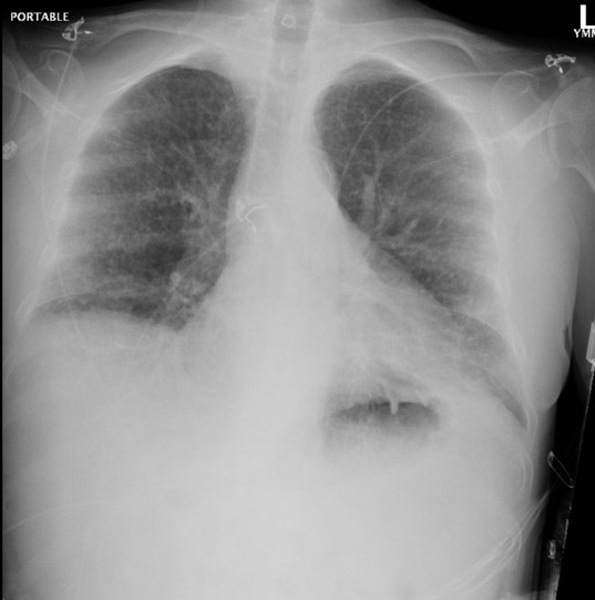
Radiography

Figure 1. Admission portable chest x-ray in the emergency department. To view Figure 1 in an enlarged, separate window click here.
The patient has a history of rheumatoid arthritis (RA). Which of the following patterns of interstitial lung disease (ILD) is most common in patients with RA? (Click on the correct answer to be directed to the second of seven pages)
- Acute eosinophilic pneumonia
- Lymphocytic interstitial pneumonitis
- Non-specific interstitial pneumonia
- Organizing pneumonitis
- Usual interstitial pneumonitis
February 2014 Pulmonary Case of the Month: Faster Is Not Always Better
Lewis Wesselius MD
Department of Pulmonary Medicine
Mayo Clinic Arizona
Scottsdale, AZ
History of Present Illness
A 56 year old woman with a history of rheumatoid arthritis (RA) for 26 years was seen as an outpatient. She has a recent history of increased cough, sputum and dyspnea.
PMH, FH, SH
She was originally from India but had lived in Singapore from 2011 to June 2013 before moving to Phoenix. In 2009, she was diagnosed with Mycobacterium avium-intracellulare (MAI) on bronchoscopy and started on azithromycin, ethambutol, and rifabutin. She was unable to tolerate rifabutin but was continued on ethambutol and azithromycin. She had been on etanercept for her RA which was held after the diagnosis of MAI. She had negative sputum cultures for MAI in September 2012 and her ethambutol and azithromycin were stopped. In May 2013 she had increased symptoms and bronchoscopy demonstrated Pseudomonas and nontuberculous mycobacterium (NTM). She was treated with cefipime/ciprofloxacin for 6 weeks prior to moving to Phoenix.
She does not smoke or drink. Her FH is noncontributory.
Medications
- Celecoxib 200 mg bid
- Gabapentin 600 bid
- Methotrexate 15 mg weekly
- Prednisone 5 mg daily
- Tramadol 50 mg every 4 hours prn
Physical Examination
Afebrile. SpO2 96% on room air.
Chest: scattered crackles in both lungs, no wheezes.
There were joint deformities typical of chronic RA, but otherwise the remainder of the physical exam was unremarkable.
Radiology
She brings a CT scan from 2009 (Figure 1).


Figure 1. Panels A-D: Representative static axial lung images from a thoracic CT scan performed in 2009. Lower panel: movie of selected lung images from the thoracic CT scan performed in 2009.
What should be done next? (Click on correct answer to move to next panel)
Reference as: Wesselius LJ. February 2014 pulmonary case of the month: faster is not always better. Southwest J Pulm Crit Care. 2014:8(2):74-8. doi: http://dx.doi.org/10.13175/swjpcc168-13 PDF
May 2012 Pulmonary Case of the Month: Things Are Not Always as They Seem
Lewis J. Wesselius, MD
Pulmonary Medicine
Mayo Clinic Arizona
Scottsdale, AZ
History of Present Illness
A 69 year old woman was seen for side effects of corticosteroids. She is a winter visitor to Arizona. She was hospitalized in March 2008 with increased dyspnea and cough and had an abnormal CT chest. A VATS lung biopsy was performed. The pathology of the lung biopsy interpreted as bronchiolitis obliterans. She was started on prednisone 60 mg/day.
Subsequently, she returned to Minnesota and was seen by rheumatologist with a diagnosis made of possible rheumatoid arthritis. She was treated with methotrexate (12.5 mg weekly) and continued prednisone at 20 mg/day from 2008 to 2011. At that time a question was raised of methotrexate lung toxicity and it was stopped but she continued on prednisone 20 to 40 mg/day. She is currently having issues with steroid side effects and seen for a second opinion.
PMH, SH and FH
She has a history of knee and other joint pains. She had knee replacement surgery in Jan 2008 with worsening of her dyspnea and cough. She has a history of diabetes which was apparently induced by the corticosteroids. Her current medications include prednisone 20 mg/day, insulin, metformin, lovastatin. She is a former smoker with 25 pack-years but quit 25 years ago. She has no family history of lung disease.
Physical Examination
She was an obese woman appearing somewhat Cushingoid in no acute distress. On chest auscultation she had diminished breath sounds but no crackles or wheezes. Examination of her joints revealed no abnormalities. The remainder of her physical examination was normal.
Chest X-ray
Her chest x-ray was interpreted as normal.
Which of the following are indicated?
- Pulmonary function testing
- Pulmonary CT scanning
- Rheumatologic evaluation
- Repeat of open lung biopsy
- All of the above
Reference as: Wesselius LJ. May 2012 pulmonary case of the month: things are not always as they seem. Southwest J Pulm Crit Care 2012;4:142-8. (Click here for a PDF version of the case)