
Pulmonary
The Southwest Journal of Pulmonary and Critical Care publishes articles broadly related to pulmonary medicine including thoracic surgery, transplantation, airways disease, pediatric pulmonology, anesthesiolgy, pharmacology, nursing and more. Manuscripts may be either basic or clinical original investigations or review articles. Potential authors of review articles are encouraged to contact the editors before submission, however, unsolicited review articles will be considered.
September 2024 Pulmonary Case of the Month: An Ounce of Prevention Caused a Pound of Disease
University of Nebraska Medical Center
Omaha, NE USA
History of Present Illness
A 55-year-old woman is self-referred for dizziness, fatigue, and difficulty concentrating. She was well until 2 months prior to this visit. She says she feels like she is in a “fog”. She also complains of a “tight chest”.
PMH, SH, and FH
She has a past medical history of hypertension and presently takes metoprolol. She has had a tubal ligation and a breast lumpectomy in the past. There is a questionable history of a positive Cardiolite nuclear stress test.
She is divorced and lives alone in a small town in Iowa. She does not smoke, drink to excess or used illicit drugs.
She has worked assembling bird houses for 20 years. She attributes her problems to a workplace exposure because she seems worse when opens the large shipping containers with the birdhouse parts. Although she worked 20 years previously without problems, her symptoms began 2 months ago after her company merged with a Chinese company. The wooden pieces are manufactured in China and the pieces are shipped to the US for assembly.
Her family history is unremarkable.
Physical Examination
Her physical examination is unremarkable.
Which of the following are indicated for further workup?
- Cardiology referral
- Neuropsychological testing
- Pulmonary function testing (PFTs)
- 1 and 3
- All of the above
June 2023 Pulmonary Case of the Month: An Invisible Disease
Pulmonary Department
Mayo Clinic Arizona
Scottsdale, AZ USA
History of Present Illness
A 78-year-old man presented to the Emergency Department on April 7 for shortness of breath and weakness over the last 2 weeks. He was in good health prior to an outside hospitalization March 29-April 3 for pneumonia and a possible non-ST-elevation myocardial infarction (elevated troponins). He had a bronchoscopy during his recent outside hospitalization without specific pathogen identified but was treated with antibiotics and discharged on levofloxacin. Since his hospital discharge 4 days previously he feels weaker and increasingly short of breath. He is short of breath even walking around his home. He denies fever or a productive cough.
Past Medical History, Family History and Social History
- Atrial fibrillation, s/p ablation. On Eliquis.
- Prior renal cell carcinoma, s/p resection, no recurrence
- DM Type 2
- GERD
- OSA
- Essential tremor
- Never smoked
Medications
- Apixaban
- Aspirin
- Atorvastatin
- Flecanide
- Insulin
- Levofloxacin
- Lisinopril
- Pantoprazole
- Tamsulosin
Physical Examination
- General: The patient looks comfortable and is in no distress
- Vital Signs: BP 110/62 O2 Sat 94% on room air
- CVS: Heart sounds are regular
- Lungs: Clear to auscultation
- Abdomen: Soft, nontender, bowel sounds present
- Extremities: No edema
- Neuro: Alert and oriented
- Skin: Warm and dry, no rashes
Chest X-ray
A portable chest X-ray was performed (Figure 1).

Figure 1. Portable chest X-ray obtained in the emergency department.
Which of the following should be done next? Click on the correct answer to be directed to the second of six pages)
February 2023 Pulmonary Case of the Month: SCID-ing to a Diagnosis
Pulmonary Department
Mayo Clinic Arizona
Scottsdale, AZ USA
History of Present Illness
A 40-year-old man was referred for management of respiratory symptoms of cough, sputum production and shortness of breath. He has a history of respiratory infections that began in early childhood. Sputum cultures were positive for Pseudomonas. He is currently using oxygen at night and occasionally during the day.
Past Medical History, Family History and Social History
- Childhood diagnosis of asthma.
- Multiple colds and pneumonias in the past.
- No family history of a similar problem.
- He has never smoked.
- Denies any occupational exposure.
Physical Examination
- Vital Signs: O2 Sat 88% on RA
- Chest: diminished breath sounds, no wheezes
- Heart: regular rate and rhythm without murmur
- Extremities: mild clubbing present, no edema
Pulmonary Function Testing
Pulmonary function testing (PFTs) was performed with results as below (Figure 1).
Figure 1. Pulmonary function testing.

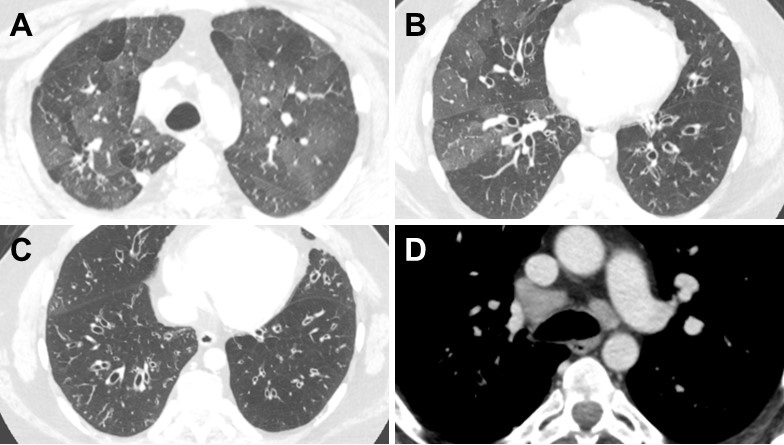
Thoracic CT Scan
A thoracic CT was performed (Figure 2).

Figure 2. Representative images from the thoracic CT in lung windows (A-C) and soft tissue windows (D). To view Figure 2 in a separate enlarged window click here
Which of the following is/are true? (Click on the correct answer to be directed to the second of six pages)
- PFTs show severe obstructive disease
- The thoracic CT shows a normal mediastinum
- Bronchiectasis is shown in the CT scan lung windows
- 1 and 3
- All of the above
December 2021 Pulmonary Case of the Month: Interstitial Lung Disease with Red Knuckles
Department of Pulmonary Medicine
Mayo Clinic Arizona
Scottsdale, AZ USA
History of Present Illness
A 56-year-old man was referred for a second opinion on recent onset of diffuse parenchymal lung disease. He had started noting mild dyspnea with yard work approximately in March 2021. His symptoms progressed over the next month with increasing shortness of breath and some fever. He presented to outside emergency department on April 17, 2021 and chest CT showing patchy ground-glass opacities with some areas of irregular consolidation (Figure 1).

Figure 1. Representative images from the thoracic CT in lung windows from outside emergency room visit.
He was subsequently seen by an outside pulmonologist and started empirically on prednisone (50 mg/day). An outside lung biopsy had been performed which showed nonspecific interstitial pneumonitis. There was some improvement in his symptoms and his prednisone dose was reduced to 20 mg/day; however, his symptoms subsequently worsened with saturations noted to drop to 85% with any ambulation. He also had swelling of his left face and a biopsy of the parotid gland with the findings suggestive of malignancy, possibly melanoma.
What should be done at this time? (Click on the correct answer to be directed to the second of seven pages)
- History and physical examination
- Repeat the open lung biopsy
- Repeat the parotid biopsy
- 1 and 3
- All of the above
A Case Series of Electronic or Vaping Induced Lung Injury
Ronald Ferrer Espinosa DO1
Abdirahman Hussein MD2
Matthew Sehring DO1
Mohamad Rachid MD1
Ryan Dunn MD1
Deepak Taneja MD1
Department of Pulmonary and Critical Care Medicine1
Department of Internal Medicine2
University of Illinois College of Medicine at Peoria
Peoria, Illinois
Abstract
Introduction: Since their introduction, electronic cigarette use has increased and was even proposed as an alternative to traditional tobacco use. Recently, a series of patients with acute respiratory failure due electronic cigarette, or vaping, associated lung injury (EVALI) in 2019 has been described which has largely been attributed to tetrahydrocannabinol (THC) containing vaporizer itself, as well as vitamin E acetate. Several case series have been published regarding the acute presentation, diagnosis and management. In addition to diagnosis and management of EVALI, we sought to describe potential long-term effects of lung parenchyma in these patients.
Methods: A retrospective review was performed on 16 patients with clinically diagnosed EVALI at OSF St Francis Medical Center between August 01 2019 and February 1 2020. Relevant demographic and clinical data were collected in patients diagnosed with EVALI.
Results: Of the 16 patients in the study the median age (IQR) age was 25.25 (20-29) and 94% were male. The predominant presenting symptoms were dyspnea (94%), cough (56%), nausea 63%), vomiting (63%), abdominal pain (50%), diarrhea (50%), and fever (63%). 2 (13%) patients required endotracheal intubation. Common features of computerized tomography (CT) scan were bilateral diffuse ground glass opacity (93%), septal thickening (53%), and subpleural sparing (47%). Bronchoalveolar lavage (BAL) was obtained in 3 patients and all demonstrated neutrophil predominance of 69% (56-90). One BAL was significant for hemosiderin laden macrophages. Post hospital follow up pulmonary function tests were obtained in 3 and 2 of these were significant for obstructive lung disease.
Conclusions: In this case series of patients diagnosed with vaping associated lung injury, obstructive lung disease may be seen on pulmonary function testing and surveillance of these patients should occur regardless of duration.
Keywords: CT scan, EVALI, bronchoalveolar lavage, electronic cigarette, pulmonary function testing, respiratory failure, tetrahydrocannabinol, vaping, vaping associated lung injury, vitamin E,
Introduction
The first cases of vaping associated lung injury (EVALI) were reported in Wisconsin and Illinois in the summer of 2019 which reached its peak in the fall of 2019 (1). This sudden epidemic of respiratory failure in patients who used tetrahydrocannabinol (THC) containing vaporizing devices lead the Food and Drug Administration (FDA), Center for Disease Control (CDC) and local public health departments to initiate investigations and research into the causative mechanisms of this disease. Currently, it is postulated that vitamin E acetate plays a role in the pathogenesis of VALI, as this substance was found in samples of vaping cartridges and in the bronchoalveolar fluid of patients with the disease (2,3). These pathological pathways are still being elucidated. Little is known about the long-term damage to the respiratory system in patients with EVALI. In this study, we sought to describe the diagnostic commonalities in patients with EVALI and describe potential long-term complications.
Methods
The study was a retrospective cohort analysis. Institutional Review Board (IRB) approval (1593746-1) was obtained through the University of Illinois College of Medicine at Peoria IRB. Data was collected for consecutive patients over 18 years of age who were diagnosed with EVALI between the dates of August 1, 2019 and February 1, 2020. The diagnosis of EVALI was consistent with the outbreak case surveillance definition. A confirmed case required the following to satisfy criteria: e-cigarette or dabbing in 90 days before symptom onset, pulmonary infiltrate, absence of infection, and no evidence of alternative plausible causes. A presumptive case definition included the above definition except for possibility of another cause of the patient’s symptoms such as infection. Data were extracted from the electronic medical record. The recorded data included the following: age, gender, co-morbidities, tobacco and electronic cigarette use history, need for endotracheal intubation, symptoms on presentation to the emergency department, computerized tomography findings, pulmonary function test values, bronchoalveolar lavage fluid studies, and discharge treatment plans. Obstructive lung disease is based on the American Thoracic Society/European Respiratory Society criteria that recommends the fifth percentile of the distribution in a population of healthy lifelong nonsmokers as the lower limit of normal. One patient described in this study has been previously described (4). Continuous variables are presented at median and interquartile range (IQR) with 95% CI. Categorical variables are described as number of patients (percentage).
Results
From August 1, 2019 to February 1, 2020, a total of 16 patients with either confirmed or presumptive vaping associated lung injury were reviewed. Table 1 shows the demographic data obtained from these patients.

The median age was 25.25 (IQR, 20-29) and the majority of patients were male 94% (n=15). Only 13% (n=2) of patients had previously diagnosed lung conditions, both of which were asthma. Of the reported THC brands, Dank© was the most commonly reported occurring in 25% (n=4) of patients (Table 2).

However, 50% (n=8) of THC products were not clearly stated in the patient’s medical record. Tobacco cigarette and tobacco electronic cigarette use were also documented, occurring in 63% (n=10) and 44% (n=7) of patients, respectively. Of those reporting tobacco use, the median pack years was 1.5 (IQR, 0.5-4.25). 7 patients reported no prior tobacco use. 13% (n=2) of patients required endotracheal intubation
Patients' symptoms are summarized in Table 3.

Any respiratory symptom occurred in 94% (n=15) of patients which included dyspnea 94% (n=15), cough 56% (n=9), chest pain 25% (n=4), and hemoptysis 19% (n=3). Abdominal symptoms were common and occurred in 75% (n=12) of patients. The most common gastrointestinal symptom were both nausea and vomiting 63% (n=10). These were followed by abdominal pain and diarrhea 50% (n=8). Fever was the most common constitutional symptom occurring in 63% (n=10) of patients. Less common constitutional symptoms included fatigue and chills both of which occurred in 13% (n=2) of patients.
Chest computerized tomography (CT) scans were available in 94% (n=15) of patients. The findings are summarized in Table 4.

The most common radiographic feature was bilateral diffuse ground glass opacity (GGO), which occurred in 93% (n=14) of patients. Septal thickening and subpleural sparing were the next most common radiographic findings, occurring in 53% (n=8) and 47% (n=7) patients, respectively. Less common features that were described on chest CT scan were centrilobular nodular consolidations occurring in 27% (n=4) and reverse haloing occurring in 7% (n=1, Figure 1).

Figure 1. Reverse halo sign in a patient with EVALI.
Three patients diagnosed with EVALI underwent bronchoscopy with bronchoalveolar lavage which are summarized in table 5.

Bronchoscopy was performed when the outbreak case definition was not met or there was a clinical concern for a secondary pathological process. The “typical” bronchoalveolar lavage (BAL) differential was neutrophilic predominant (56%-90%) with elevated macrophage and/or monocyte counts (4-19%, 0-23%). The lymphocyte count was typically low (0-4%) as well as the eosinophil count (0-1%). All the microbiologic data from these lavage samples were negative and included the following tests: aerobic and anaerobic bacterial cultures, fungal cultures, silver stain, acid fast bacilli smear and culture, varicella zoster polymerase chain reaction (PCR), histoplasma antigen and galactomannan. The results of cytology were different in all three BAL samples and were significant fore alveolar macrophages, atypical glandular cells and hemosiderin laden macrophages. At the time of bronchoscopy, only 1 or 3 patients was on or received systemic steroids therapy.
A few complications occurred in our cohort which included pneumothorax, pneumorrhachis, pulmonary embolism and Takutsubo cardiomyopathy. The case of Takusubo cardiomyopathy has been reported in the literature (4). The rate of pneumothorax was 13% (n=2). The pneumorrhachis 6% (n=1) occurred in a patient with pneumothoraces. Pulmonary embolism was only seen in 1 patient.
The discharge treatment course involved mainly systemic corticosteroids which were given in 81% (n=13) of patients. Antibiotics were continued at discharge in a minority of patients 38% (n=6). The majority of patients (94%) recovered and were asymptomatic at a later clinic visit. One patient remained symptomatic with persistent dyspnea.
Full pulmonary function tests were obtained in 3 patients (Table 6).

The timing of the pulmonary function tests occurred 3-4 weeks post hospital discharge. THC vape duration for these patients ranged from 4-12 weeks and tobacco pack years ranged from 0-2. Forced expiratory volume in 1 second to forced vital capacity (FEV1/FVC) ratios were 70%, 71%, and 86%. FEV1% ranged from 70 to 119%. FVC ranged from 81% to 119%. The total lung capacity in these three patients ranged from 85% to 109%. Diffusing capacity of carbon monoxide (DLCO) ranged from 60% to 100%. The flow volume loops of two patients demonstrate coving of the expiratory limb (Figure 2).

Pre-EVALI pulmonary function testing was not available for review in any of the patients.
Discussion
In this case series of patients with vaping associated lung injury of 16 patients in Central Illinois from August 1 2019 through February 1, 2020 the majority were younger men with respiratory and gastrointestinal symptoms. The symptomatology was consistent with previously published data (1,5,6,7). The exact pathophysiologic mechanisms implicated in vaping associated lung injury are currently still under investigation, but vitamin E acetate has recently been implicated possibly through the transition of tocopherols induced transition of phosphatidiylcholines from gel to liquid crystalline, causing surfactant dysfunction (2). The same authors proposed an alternative mechanism whereby ketene, created while heating e-cigarette products, causes direct lung irritation. However, many branded vaping products are not commercially produced and the heterogenous ingredients such as propylene glycol and other flavoring ingredients in these products may reflect its informal creation, which may influence the development of different disease mechanisms, clinical phenotypes and imaging findings (8,9).
A growing body of EVALI cases demonstrate predominant features on computerized tomography which include basilar predominant centrilobular nodular ground glass opacities and ground glass opacities with subpleural sparing. An initial study proposed four imaging patterns which included acute eosinophilic pneumonia, diffuse alveolar damage, organizing pneumonia and lipoid pneumonia and predicted the dominant CT scan findings (10). Later in a small case series of pediatric patients, centrilobular ground glass opacities were present in 92% and ground glass opacities with subpleural sparing were present in 75% of patients (11). The importance of recognizing these patterns is essential, especially in the adolescent and young adult populations where disclosure of medical history may prove to be difficult. Our findings support the cases in literature, with 93% of patients having bilateral diffuse basilar predominant GGO (93%), some associated with subpleural sparing (47%) and to a lesser degree centrilobular nodular consolidation (27%).
The role of bronchoscopy in patients with vaping associated lung injury has waned with as the characterization of clinical and radiographic findings has matured. Importantly, there is no finding on bronchoscopy that is specific for diagnosis of EVALI. Recently, the role of bronchoscopy with bronchoalveolar lavage (BAL) has been suggested to be performed in cases with a high pretest probability of EVALI with atypical features that cannot be attributed to vaping (5). Lipid laden macrophages (LLM) are a non-specific finding in many different illnesses such as infection, aspiration, drug reactions, lipoid pneumonia, pulmonary alveolar proteinosis and autoimmune disorders, but the absence of LLM, argued by Aberegg, would be an atypical finding in EVALI (5,12). Our BAL fluid differentials are consistent with the published data, with elevations in neutrophils and/or monocytes and macrophages, with scant lymphocytes and eosinophils. In our institution, staining for lipid laden macrophages was not routinely performed. However, in one of our cytologic BAL samples hemosiderin macrophages were identified. The inconsistent cytologic findings on BAL in our series supports the notion of heterogenous underlying pathophysiologic mechanisms involved in EVALI.
The long-term effects of EVALI on lung parenchyma are unknown. Prior studies evaluating the effects of electronic cigarette use prior to the EVALI epidemic have suggested airway hyper responsiveness and an obstructive pattern on spirometry. In mice, it has been previously demonstrated that aerosolized nicotine induced airway hyperreactivity (13). A human study of 30 electronic cigarette users with at least 6 months of use compared with matched controls, demonstrated PFTs consistent with peripheral obstruction (14). A different study comparing vaping asthmatics versus healthy controls demonstrated acute declines in the FEV1/FVC ratio (15). Additionally, a few recent reports of pulmonary function testing have been documented in patients diagnosed with EVALI in late 2019 and early 2020. One study of intubated adults reported one follow-up PFT that was significant for only a low DLCO (16). A case series of pediatric patients describes two patients with 6 week follow up PFT’s that were significant for obstructive lung disease, and one of these patients had a low DLCO (11). Our study contributes 3 full PFT’s in adult patients diagnosed with EVALI performed at either 3 or 4 weeks following hospital discharge. Two of these PFT’s demonstrated obstructive lung disease, of which one had a low DLCO. The duration of vaping in these two abnormal cases were 8 and 12 weeks, and the pack years of tobacco use were 2 and 1.25 years. This small data set suggests that patients who participate in electronic cigarette use, or vaping, may be at higher risk for developing obstructive lung disease regardless of the duration of use. It is also important to take into account that a fixed FEV1/FVC ratio may underestimate young patients with obstructive lung disease (17). Using the lower limit of normal in this younger patient population is likely to be more sensitive in detecting obstructive physiology. It is plausible that vaping may cause an accelerated obstructive lung pattern due to the many potential pathways for lung injury. Full pulmonary function testing in any patient who was diagnosed with EVALI or continues to use electronic cigarettes, or vaping, products should be considered.
This study has several limitations. First, this was a retrospective study of patients at a single center in one geographic location. Second, our sample size was relatively small. Third, our clinical follow up rate was only 50%, of which only 37% completed PFT’s.
Conclusions
In our case series of 16 patients, predominantly male, diagnosed with EVALI there was a high incidence of gastrointestinal symptoms on presentation and follow up pulmonary function tests suggested there may be an increased risk for obstructive lung disease. Avoidance of vaping products, especially “Dank” and other similarly formulated products, is strongly recommended.
References
- Layden JE, Ghinai I, Pray I, et al. Pulmonary Illness Related to E-Cigarette Use in Illinois and Wisconsin - Final Report. N Engl J Med. 2020 Mar 5;382(10):903-916. [CrossRef] [PubMed]
- Blount BC, Karwowski MP, Shields PG, et al. Vitamin E Acetate in Bronchoalveolar-Lavage Fluid Associated with EVALI. N Engl J Med. 2020 Feb 20;382(8):697-705. [CrossRef] [PubMed]
- Taylor J, Wiens T, Peterson J, et al. Characteristics of E-cigarette, or Vaping, Products Used by Patients with 18 and 2019. MMWR Morb Mortal Wkly Rep. 2019 Nov 29;68(47):1096-1100. [CrossRef] [PubMed]
- Rachid M, Yin J, Mier N, et al. Reversed Takutsubo Cardiomyopathy in a young patient with vaping induced acute lung injury. ATS International Conference Abstract Presentation. 2020 May 15-20. Philadelphia, PA. Abstract nr A1839.
- Aberegg SK, Maddock SD, Blagev DP, Callahan SJ. Diagnosis of EVALI: General Approach and the Role of Bronchoscopy. Chest. 2020 Aug;158(2):820-827. [CrossRef] [PubMed]
- Blagev DP, Harris D, Dunn AC, Guidry DW, Grissom CK, Lanspa MJ. Clinical presentation, treatment, and short-term outcomes of lung injury associated with e-cigarettes or vaping: a prospective observational cohort study. Lancet. 2019 Dec 7;394(10214):2073-2083. [CrossRef] [PubMed]
- Kalininskiy A, Bach CT, Nacca NE, et al. E-cigarette, or vaping, product use associated lung injury (EVALI): case series and diagnostic approach. Lancet Respir Med. 2019 Dec;7(12):1017-1026. [CrossRef] [PubMed]
- Heinzerling A, Armatas C, Karmarkar E, et al. Severe Lung Injury Associated With Use of e-Cigarette, or Vaping, Products-California, 2019. JAMA Intern Med. 2020 Jun 1;180(6):861-869. [CrossRef] [PubMed]
- Puebla Neira D, Tambra S, Bhasin V, Nawgiri R, Duarte AG. Discordant bilateral bronchoalveolar lavage findings in a patient with acute eosinophilic pneumonia associated with counterfeit tetrahydrocannabinol oil vaping. Respir Med Case Rep. 2020 Feb 3;29:101015. [CrossRef] [PubMed]
- Henry TS, Kanne JP, Kligerman SJ. Imaging of Vaping-Associated Lung Disease. N Engl J Med. 2019 Oct 10;381(15):1486-1487. [CrossRef] [PubMed]
- Thakrar PD, Boyd KP, Swanson CP, Wideburg E, Kumbhar SS. E-cigarette, or vaping, product use-associated lung injury in adolescents: a review of imaging features. Pediatr Radiol. 2020 Mar;50(3):338-344. [CrossRef] [PubMed]
- Larsen BT, Butt YM, Smith ML. More on the Pathology of Vaping-Associated Lung Injury. Reply. N Engl J Med. 2020 Jan 23;382(4):388-390. [CrossRef] [PubMed]
- Larcombe AN, Janka MA, Mullins BJ, Berry LJ, Bredin A, Franklin PJ. The effects of electronic cigarette aerosol exposure on inflammation and lung function in mice. Am J Physiol Lung Cell Mol Physiol. 2017 Jul 1;313(1):L67-L79. [CrossRef] [PubMed]
- Meo SA, Ansary MA, Barayan FR, Almusallam AS, Almehaid AM, Alarifi NS, Alsohaibani TA, Zia I. Electronic Cigarettes: Impact on Lung Function and Fractional Exhaled Nitric Oxide Among Healthy Adults. Am J Mens Health. 2019 Jan-Feb;13(1):1557988318806073. [CrossRef] [PubMed]
- Kotoulas SC, Pataka A, Domvri K, et al. Acute effects of e-cigarette vaping on pulmonary function and airway inflammation in healthy individuals and in patients with asthma. Respirology. 2020 Oct;25(10):1037-1045. [CrossRef] [PubMed]
- Choe J, Chen P, Falk JA, Nguyen L, Ng D, Parimon T, Ghandehari S. A Case Series of Vaping-Associated Lung Injury Requiring Mechanical Ventilation. Crit Care Explor. 2020 Jan 29;2(1):e0079. [CrossRef] [PubMed]
- Cerveri I, Corsico AG, Accordini S, et al. Underestimation of airflow obstruction among young adults using FEV1/FVC <70% as a fixed cut-off: a longitudinal evaluation of clinical and functional outcomes. Thorax. 2008 Dec;63(12):1040-5. [CrossRef] [PubMed]
Cite as: Espinosa RF, Hussein A, Sehring M, Rachid M, Dunn R, Taneja D. A Case Series of Electronic or Vaping Induced Lung Injury. Southwest J Pulm Crit Care. 2021;23(2):62-9. doi: https://doi.org/10.13175/swjpcc032-21 PDF
March 2020 Pulmonary Case of the Month: Where You Look Is Important
Richard A. Robbins, MD
Anselmo Garcia, MD
Arizona Chest and Sleep Medicine
Phoenix, AZ USA
History of Present Illness
A 47-year-old woman was seen for the first time in our clinic. She had approximately a two-year history of gradually increasing shortness of breath to the point where she could only climb one flight of stairs. In addition, she has a history of a cough sometimes productive and sometimes nonproductive. She did hear herself wheeze intermittently.
PMH, SH, and FH
She has a past medical history of gastroesophageal reflux disease (GERD). She was a nonsmoker and had no occupational exposure. Her aunt has a history of asthma.
Physical Examination
Her physical examination was normal and her lungs were clear.
Which of the following is appropriate at this time?
- Reassurance
- Treat empirically for post-nasal drip
- Treat empirically with albuterol
- Treat empirically with omeprazole
- None of the above
Cite as: Robbins RA, Garcia A. March 2020 pulmonary case of the month: where you look is important. Southwest J Pulm Crit Care. 2020;20(3):76-83. doi: https://doi.org/10.13175/swjpcc013-20 PDF
June 2019 Pulmonary Case of the Month: Try, Try Again
Lewis J. Wesselius, MD
Department of Pulmonary Medicine
Mayo Clinic Arizona
Scottsdale, AZ USA
History of Present Illness
A 53-year-old woman from presented with a 3-year history of shortness of breath. She was diagnosed with pneumonia in 2016, but even after treatment with antibiotics, continued to require supplemental oxygen. A CT-guided biopsy of a lung nodule was performed but there were no diagnostic findings. A surgical lung biopsy at another hospital was done but the report is unavailable. She had been diagnosed with possible scleroderma and treated with mycophenolate for 3 months and then azathioprine.
Past Medical History, Social History and Family History
Aside from her history as in the HPI she has a remarkably negative past medical history. She does not smoke. Family history is noncontributory.
Physical Examination
- HEENT: negative
- Chest: Fine crackles at both lung bases
- Cardiovascular: regular rhythm, no murmur
- Skin: skin thickening on fingers and distal forearms, but not elsewhere. No pitting, ulcerations or calcinosis
Radiology
A chest x-ray was performed (Figure 1).

Figure 1. PA chest radiography done on presentation.
Which of the following should be done? (Click on the correct answer to be directed to the second of six pages)
- Obtain previous radiography and biopsy reports
- Pulmonary function testing
- Thoracic CT scan
- 1 and 3
- All of the above
Cite as: Wesselius LJ. June 2019 pulmonary case of the month: Try, try again. Southwest J Pulm Crit Care. 2019;18(6):144-51. doi: https://doi.org/10.13175/swjpcc026-19 PDF
September 2018 Pulmonary Case of the Month: Lung Cysts
Lewis J. Wesselius, MD
Department of Pulmonary Medicine
Mayo Clinic Arizona
Scottsdale, AZ USA
Pulmonary Case of the Month CME Information
Completion of an evaluation form is required to receive credit and a link is provided on the last page of the activity.
0.50 AMA PRA Category 1 Credit(s)™
Estimated time to complete this activity: 0.50 hours
Lead Author(s): Lewis J. Wesselius, MD. All Faculty, CME Planning Committee Members, and the CME Office Reviewers have disclosed that they do not have any relevant financial relationships with commercial interests that would constitute a conflict of interest concerning this CME activity.
Learning Objectives: As a result of completing this activity, participants will be better able to:
- Interpret and identify clinical practices supported by the highest quality available evidence.
- Establish the optimal evaluation leading to a correct diagnosis for patients with pulmonary, critical care and sleep disorders.
- Translate the most current clinical information into the delivery of high quality care for patients.
- Integrate new treatment options for patients with pulmonary, critical care and sleep related disorders.
Learning Format: Case-based, interactive online course, including mandatory assessment questions (number of questions varies by case). Please also read the Technical Requirements.
CME Sponsor: The University of Arizona College of Medicine-Tucson
Current Approval Period: January 1, 2017-December 31, 2018
Financial Support Received: None
History of Present Illness
A 67-year-old woman was referred for mild shortness of breath for several years, but worse since January 2018. She has dyspnea on exertion after 1 block. An outside chest x-ray, electrocardiogram and echocardiogram are reported as normal. She was begun on prednisone at 40 mg/day and her symptoms improved. However, her symptoms worsened when the dose tapered to 5 mg/day. She gained 35 pounds while on the prednisone and tried a steroid inhaler therapy without benefit. She is still dyspneic after 1 block of exertion.
Past Medical History, Social History, Family History
- Her past medical history was only positive for gastroesophageal reflux for which she takes ranitidine and hypertension for which she takes lisinopril.
- She was a life-long nonsmoker.
- There was no occupational history, hot tub or bird exposures.
- Family history is noncontributory.
Physical Examination
- Her SpO2 was 94% on room air.
- Chest: few crackles noted at right base.
- Cardiovascular: regular rate and rhythm without a murmur.
- Extremities: no edema or clubbing.
Which of the following should be done at this time? (Click on the correct answer to be directed to the second of eight pages)
- Measure her SpO2 after exercise
- Reassure the patient the patient that she has hysterical dyspnea
- Pulmonary function testing
- 1 and 3
- All of the above
Cite as: Wesselius LJ. September 2018 pulmonary case of the month: lung cysts. Southwest J Pulm Crit Care. 2018;17(3):85-92. doi: https://doi.org/10.13175/swjpcc101-18 PDF
April 2014 Pulmonary Case of the Month: DIP-What?
Lewis Wesselius MD
Department of Pulmonary Medicine
Mayo Clinic Arizona
Scottsdale, AZ
History of Present Illness
A 53 year old woman from Indiana was seen who had a history of nonproductive cough for several years. She had a prior diagnosis of asthma but continued to have cough despite asthma treatment. She was also treated for gastroesophageal reflux and had a Nissen fundoplication. This resolved in some improvement in the cough. In May 2013 she noted increasing dyspnea on exertion.
An echocardiogram was performed which was notable for a 16% left ventricular ejection fraction. A thoracic CT demonstrated some nodules and a question was raised of sarcoidosis. She was admitted to a hospital in Indiana and had a biventricular pacemaker placed. Bronchoscopy with transbronchial biopsy was performed with no diagnostic findings. No granulomas were seen on the biopsy. Bronchoalveolar lavage showed a CD4/CD8 ration of 0.84. Optic nerve swelling was noted at that time. Due to the cardiac, pulmonary, and optic nerve findings a clinical diagnosis of sarcoidosis with a dilated cardiomyopathy was made and she was treated with prednisone initially, then a combination of prednisone and methotrexate.
PMH, FH, SH
Her past medical history was as above and family history was noncontributory. She does not smoke or drink.
Medications
- Methotrexate 15 mg weekly
- Prednisone 5 mg daily
- Furosemide 40 mg daily
- Potassium chloride 20 meq daily
Physical Examination
Afebrile. SpO2 96% on room air. The physical exam was unremarkable.
Which of the following should be performed at this time?
Reference as: Wesselius LJ. April 2014 pulmonary case of the month: DIP-what? Southwest J Pulm Crit Care. 2014;8(4):195-203. doi: http://dx.doi.org/10.13175/swjpcc024-14 PDF