
Pulmonary
The Southwest Journal of Pulmonary and Critical Care publishes articles broadly related to pulmonary medicine including thoracic surgery, transplantation, airways disease, pediatric pulmonology, anesthesiolgy, pharmacology, nursing and more. Manuscripts may be either basic or clinical original investigations or review articles. Potential authors of review articles are encouraged to contact the editors before submission, however, unsolicited review articles will be considered.
December 2023 Pulmonary Case of the Month: A Budding Pneumonia
Sarah Medrek, MD1
Michael Reyes, MD2
Brannon Raney, MD3
Section of 1Pulmonary, Critical Care, and Sleep Medicine, 2Pathology, and 3Infectious Disease
VA Albuquerque Health System
Albuquerque, NM USA
History of Present Illness
A 70-year-old man with a history of seropositive rheumatoid arthritis previously well controlled on hydroxychloroquine, methotrexate, and adalimumab was admitted to the hospital with 3 weeks of progressively worsening fatigue, night sweats, chills, and malaise. He did not describe new or worsening cough, shortness of breath, or sputum production. On the day of admission, he had intense nausea and vomiting.
PMH, SH, and FH
Prior to this admission, he was followed in Pulmonary Clinic for asymptomatic mild basilar fibrosis thought to be related to his rheumatoid arthritis and paraseptal emphysema related to prior smoking which was largely stable and unchanged over the previous two years. Previously, he smoked cigarettes at ½ pack per day for about 30 years and quit about 15 years ago. He denied any recent travel and was retired from the last 15 years from being a meat butcher. FH is noncontributory.
Physical Examination
On examination the day after admission from the ER, the patient’s temperature was 37.6C. His pulse was 79 bpm, blood pressure was 142/65 mmHg, and pulse oximetry revealed a saturation of 92% with 2 LPM nasal cannula of O2. He appeared generally weak, but alert. Pulmonary exam was unrevealing as was cardiac exam. He did not have cyanosis, clubbing, delayed capillary refill, or peripheral edema.
Laboratory
Initial blood work showed a WBC count of 7500/µL, hemoglobin level of 9.6 gm/dl, serum blood urea nitrogen of 36 gm/dl, serum creatinine of 2.49 g/dl, and serum calcium that was elevated at 12.3 mg/dl. A T-spot was obtained and was negative. Blood and sputum cultures were obtained and negative.
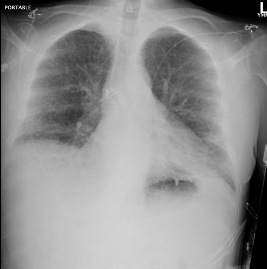
Radiography

Figure 1. Admission portable chest x-ray in the emergency department. To view Figure 1 in an enlarged, separate window click here.
The patient has a history of rheumatoid arthritis (RA). Which of the following patterns of interstitial lung disease (ILD) is most common in patients with RA? (Click on the correct answer to be directed to the second of seven pages)
- Acute eosinophilic pneumonia
- Lymphocytic interstitial pneumonitis
- Non-specific interstitial pneumonia
- Organizing pneumonitis
- Usual interstitial pneumonitis
A Summary of Outpatient Recommendations for COVID-19 Patients and Providers December 9, 2021
Richard A. Robbins MD1
Stephen A. Klotz MD2
1Phoenix Pulmonary and Critical Care Research and Education Foundation, Gilbert, AZ USA
2Division of Infectious Disease, Department of Medicine, University of Arizona College of Medicine, Tucson, AZ USA
We thought a follow-up to our original brief review of COVID-19 in February, 2020 might be useful. As we write this in early December 2021, we again caution that this area is rapidly changing and what is true today will likely be outdated tomorrow. We again borrowed heavily from the Centers for Disease Control (CDC) CDC website and the NIH website which have extensive discussions over numerous pages covering COVID-19. Our hope is to condense those recommendations. We do not discuss inpatient care in any detail.
COVID-19 Variants
The initial steps of coronavirus infection involve the specific binding of the coronavirus spike (S) protein to the cellular entry receptors which are normally on a cell. These include human aminopeptidase N (APN; HCoV-229E), angiotensin-converting enzyme 2 (ACE2; HCoV-NL63, SARS-CoV and SARS-CoV-2) and dipeptidyl peptidase 4 (DPP4; MERS-CoV).
All viruses, but especially simple single-stranded RNA viruses like COVID-19, constantly change through mutation resulting in new variants (1). The variants vary in severity and infectivity. The CDC, World Health Organization (WHO), and other public health organizations monitor COVID-19 for emergence of new variants. Some variants emerge and disappear while others persist.
The Delta variant causes more infections and spreads faster than the original SARS-CoV-2 strain of the virus that cause COVID-19 (2). Delta is currently the predominant variant of the virus in the United States causing over 99% of infections (2). On November 24, 2021, a new variant of SARS-CoV-2, B.1.1.529, was reported to the World Health Organization (WHO). This new variant was first detected in specimens collected on November 11, 2021 in Botswana and on November 14, 2021 in South Africa. On November 26, 2021, WHO named the B.1.1.529 Omicron and classified it as a variant of concern because of the number of mutations on the spike protein. As of this yesterday morning (12/8/21), the first Omicron case was reported in Arizona (2). Omicron is also present in California, Utah and Colorado and probably several other states since there is a lag between the presence of the virus and detection.
Early reports have suggested the Omicron variant might cause milder disease more often in children, raising hopes that the variant might be less severe than some of its predecessors (3). Dr. Müge Çevik, an infectious-disease specialist at the University of St Andrews, UK cautions, “Everyone is trying to find some data that could guide us but it’s very difficult at the moment.”
Symptoms
People with COVID-19 have had a wide range of symptoms reported – from none to severe illness (2). Symptoms may appear 2-14 days after exposure to the virus. Symptoms of flu and COVID-19 may be very similar and it may be hard to tell the difference between them based on symptoms alone. Testing may be needed to help confirm a diagnosis. COVID-19 seems to spread more easily than flu and causes more serious illnesses in some people. It can also take longer before people show symptoms and people can be contagious for longer. Despite mild symptoms, people infected with COVID-19 can still infect others.
Testing
Two types of viral tests are used: nucleic acid amplification tests and antigen tests (2). A viral test checks specimens from the nose or mouth by first reverse transcribing the RNA to DNA and then amplifying the DNA by polymerase chain reaction. COVID-19 antigen tests are designed for the rapid diagnosis of active infection primarily by detecting the nucleocapsid protein antigen of the SARS-CoV-2 virus. People who develop symptoms or have come into close contact with someone with COVID-19 should be tested 5–7 days after their last exposure or immediately if symptoms develop.
Prevention
The CDC recommends several steps for prevention of COVID-19 (2).
- Get Vaccinated. COVID-19 vaccines are protective against COVID-19, especially severe disease and death. Boosters should be administered as soon as possible.
- Wear a mask. Everyone 2 years or older who is not fully vaccinated should wear a mask in indoor public places. In general, masks are unnecessary in outdoor settings.
- However, in areas with high numbers of COVID-19 cases, consideration should be given to wearing a mask in crowded outdoor settings and for activities with close contact with others who are not fully vaccinated.
- Stay 6 feet away from others. Whenever possible, people should stay 6 feet away from others especially those who are sick. If possible, patients should be advised to maintain 6 feet between sick family members.
- Avoid crowds and poorly ventilated spaces. Crowded places like restaurants, bars, fitness centers, or movie theaters are high risk areas for spread of COVID-19. Indoor spaces that do not offer fresh air from the outdoors should be avoided.
- Test to prevent spread to others. Testing provides information about the risk of spreading COVID-19. Over-the-counter self-tests can be used at home or anywhere, are easy to use, and produce rapid results.
- Wash Hands Often. Hands should be washed often with soap and water after the patient blows their nose, coughs, sneezes, or is exposed to any public place.
- Clean and disinfect. High touch surfaces should be cleaned and disinfected regularly or as needed. This includes tables, doorknobs, light switches, countertops, handles, desks, phones, keyboards, toilets, faucets, and sinks.
Specific Groups
Any immunocompromised group or group living in close contact is at increased risk for COVID-19 infection and complications of the infection (2). This includes asthma, pregnancy, the elderly (>65 years), nearly all chronic diseases and jails or prisons.
Holidays
With Holiday gatherings here, many are concerned about COVID-19 especially with an unvaccinated relative or guest. First, the CDC recommends they get vaccinated (2). Second follow the recommendations under prevention above.
COVID-19 Patients
Patients with COVID-19, should follow the steps under prevention above (2). In addition, they stay home for 10 days after symptoms appear except to get medical care. Patients should be advised to drink fluids, take over-the-counter medications for symptomatic relief, and go to the emergency room or a physician’s office if needed, but call ahead. They should tell their close contacts that they may have been exposed to COVID-19.
COVID-19 Exposure
Patients should quarantine if you have been in close contact (within 6 feet of someone for a cumulative total of 15 minutes or more over a 24-hour period) with someone who has COVID-19, unless they are fully vaccinated (2). People who are fully vaccinated do not need to quarantine after contact with someone who had COVID-19 unless they have symptoms.
Travel
At this time patients should delay travel by bus, train, plane or ship unless fully vaccinated.
Treatment
The NIH has convened a COVID-19 Treatment Guidelines Panel (4). They recommend*:
- COVID-19 vaccination for everyone who is eligible according to the Advisory Committee on Immunization Practices (AI).
- Using one of the following anti-SARS-CoV-2 monoclonal antibodies (as post-exposure prophylaxis (PEP) for people who are at high risk of progressing to severe COVID-19:
- Bamlanivimab 700 mg plus etesevimab 1,400 mg administered as an intravenous (IV) infusion (BIII).
- Casirivimab 600 mg plus imdevimab 600 mg administered as subcutaneous injections (AI) or an IV infusion (BIII).
- Do not use hydroxychloroquine for SARS-CoV-2 PEP (AI).
- Do not use of other drugs for SARS-CoV-2 PEP, except in a clinical trial (AIII).
- Do not use any drugs for SARS-CoV-2 pre-exposure prophylaxis, except in a clinical trial (AIII).
*Rating of Recommendations: A = Strong; B = Moderate; C = Optional Rating of Evidence: I = One or more randomized trials without major limitations; IIa = Other randomized trials or subgroup analyses of randomized trials; IIb = Nonrandomized trials or observational cohort studies; III = Expert opinion
References
- Yang H, Rao Z. Structural biology of SARS-CoV-2 and implications for therapeutic development. Nat Rev Microbiol. 2021 Nov;19(11):685-700. [CrossRef] [PubMed]
- CDC. COVID-19. Available at: https://www.cdc.gov/coronavirus/2019-ncov/index.html (accessed 12-6-21).
- Callaway E, Ledford H. How bad is Omicron? What scientists know so far. Nature. 2021 Dec 2. [CrossRef] [PubMed]
- NIH. COVID-19 Treatment Guidelines. October 27, 2021. Available at: https://www.covid19treatmentguidelines.nih.gov/ (accessed 12/6/21).
Cite as: Robbins RA, Klotz SA. A Summary of Outpatient Recommendations for COVID-19 Patients and Providers December 9, 2021. Southwest J Pulm Crit Care. 2021;23(6):151-5. doi: https://doi.org/10.13175/swjpcc066-21 PDF
June 2021 Pulmonary Case of the Month: More Than a Frog in the Throat
Department of Radiology, Mayo Clinic Arizona
Phoenix, Arizona 85054
A 66-year-old woman with a history of GERD and previous renal transplant due to lithium toxicity was seen in the clinic complaining of a shortness of breath and nonproductive cough. She was on immunosuppression due to her renal transplant done about 5 months ago. These include daily trimethoprim (TMP) – sulfamethoxazole (SMX). She also had asthma and was on a long-acting bronchodilator with an inhaled corticosteroid. Because of a previous history of oropharyngeal candidiasis (thrush), she was doing nystatin swish and swallow four times a day.
Which of the following should be included in your differential diagnosis in this clinical setting? (Click on the correct answer to be directed to the second of 5 pages. Multiple guesses are allowed.)
- Candida esophagitis
- COVID-19 Infection
- Cytomegalovirus esophagitis
- Group A Streptococcus infection
- All of the above
Cite as: Gotway MB. June 2021 Pulmonary Case of the Month: More Than a Frog in the Throat. Southwest J Pulm Crit Care. 2021;22(6):109-13. doi: https://doi.org/10.13175/swjpcc017-21 PDF
September 2019 Pulmonary Case of the Month: An HIV Patient with a Fever
William P. Diehl IV, DO
Nicholas Villalobos, MD
Department of Internal Medicine
University of New Mexico
Albuquerque, NM USA
History of Present Illness
A 33-year old transgender male to female presented from human immunodeficiency virus (HIV) clinic for two months of fevers, intermittent shortness of breath, cough with blood streaked sputum, headache, and nausea. The clinic provider was concerned when labs showed up trending HIV viral load (3.3 million copies) and an absolute CD4 count of 57.
Past Medical History, Social History and Family History
The patient had a history of stage-III HIV diagnosed in 2014 on bictegravir, emtricitabine, tenofovir (Biktarvy) and latent tuberculosis (TB) diagnosed 2017 on isoniazid and B6. She is from Nicaragua and arrived in Albuquerque, NM in 2017. Social history is pertinent for sex trafficking and methamphetamine use.
Physical Examination
Upon admission, the patient’s vital signs were notable for a temperature of 39.2 degrees Celsius, blood pressure of 114/71 mmHg, oxygen saturation of 95% on room air with a respiratory rate of 18 breaths per minute. Physical exam was notable for an absence of rash, palpable lymphadenopathy or cachexia.
Which of the following should be done? (Click on the correct answer to be directed to the second of six pages)
Cite as: Diehl WP IV, Villalobos N. September 2019 pulmonary case of the month: an HIV patient with a fever. Southwest J Pulm Crit Care. 2019;19(3):87-94. doi: https://doi.org/10.13175/swjpcc056-19 PDF
First Report of Splenic Abscesses Due to Coccidioidomycosis
Shabnam Assar, MDI and Tim Kuberski, MD, FIDSA2
1Department of Medicine, Virginia Tech Carilion, Roanoke, Virginia USA
2Department of Medicine, University of Arizona School of Medicine-Phoenix,
Phoenix, Arizona USA
Abstract
Involvement of the spleen by Coccidioides is uncommon. It is usually associated only with disseminated infection and manifests as microscopic granulomas in the spleen. We report an immunosuppressed dermatomyositis patient who presented with splenic abscesses demonstrated on a computed tomography (CT) scan which was presumed to be bacterial in origin. At splenectomy the spleen was found to be filled with aggregates of spherules due to Coccidioides. Finding large splenic abscesses on CT scan due to Coccidioides has not been previously described. We offer a hypothesis for why the abscesses occurred in this unique patient.
Introduction
Involvement of the spleen by coccidioidomycosis is usually associated with disseminated disease, however the development of splenic abscesses has not been reported. Splenic involvement by coccidioidomycosis is usually manifest as microscopic miliary splenic granulomas which have been demonstrated at autopsy in patients with disseminated infection (1,2). We report an immunocompromised dermatomyositis patient who was found to have splenic abscesses due to Coccidioides spherules which were diagnosed at splenectomy.
Case Presentation
A 33-year-old Hispanic man with dermatomyositis for five years and a history of disseminated coccidioidomycosis for two years, presented to the emergency room because of left upper quadrant abdominal pain, fever and chills. Treatment of his dermatomyositis was ongoing over the previous five years and included prednisone, azathioprine and courses of intravenous immunoglobulin (IVIG) at doses of 2 g/kg (3). Treatments of his coccidioidomycosis over the previous two years included intravenous liposomal amphotericin B followed by oral fluconazole. The patient would periodically be non-compliant about taking the fluconazole and then experience relapses of his coccidioidomycosis which required additional courses of intravenous liposomal amphotericin B.
Physical Examination and Course: Admission vital signs - temperature 38.40 C; blood pressure 147/81 mmHg; heart rate 106 bpm; respiratory rate 18 breaths/minute and pulse oximetry 90% on room air. There was pigmentation of his face consistent with dermatomyositis, tenderness in the left upper quadrant and significant weakness of all extremities. He was bedridden and could barely move his arms and legs against gravity. His medications on admission were fluconazole and prednisone. An admission CT scan of the abdomen was performed because of the left upper quadrant tenderness and revealed multiple splenic abscesses (Figure 1).

Figure 1. CT scan of abdomen demonstrating splenic abscesses (arrow).
An admission urine culture grew >105 colony forming Klebsiella pneumoniae which was noted on day two of hospitalization. Blood cultures were negative. It was initially believed that the splenic abscesses were due to a Klebsiella infection because of the admitting urine culture results. Prednisone was stopped on admission and the oral fluconazole continued. Piperacillin-tazobactam was started empirically on admission. In addition, IVIG was given for a presumed dermatomyositis exacerbation. On hospital day four his abdominal pain and fevers had not improved. To avoid a splenectomy, a splenic biopsy was performed to determine the cause of the splenic abnormalities. The biopsy was consistent with a Coccidioides infection. A laparoscopic splenectomy was then preformed on hospital day seven.
The pathology on the removed spleen showed multiple necrotizing granulomatous foci containing numerous aggregated Coccidioides spherules (Figure 2).

Figure 2. Pathology of splenic abscesses demonstrating aggregated Coccidioides spherules.
Post-operatively, fluconazole was empirically replaced by voriconazole (4) and the patient was restarted on prednisone for his dermatomyositis. The fever and chills eventually resolved and he was discharged. At four months follow-up he had returned to his usual state and was encouraged to not stop taking the voriconazole.
Discussion
This patient illustrates an unusual complication of disseminated coccidioidomycosis. Prior to the advent of CT scans, splenic granulomas were described mainly at autopsy in patients with disseminated infection. Splenic involvement at autopsy was described as granulomas due to the invasion of the Coccidioides into the spleen from the blood stream. Usually there was granuloma formation described as microscopic military nodules. Reports of gross Coccidioides abscesses in the spleen have not been described.
We considered the potential reasons for the development of splenic abscesses in this unique patient. His dermatomyositis was present for about five years and the coccidioidomycosis, two years. He had received repeated doses of IVIG for flares of his dermatomyositis prior to, and after, his Coccidioides infection. Investigating his past medical history revealed that he would develop a febrile illness when off fluconazole - usually due to non-compliance. The clinical presentation was consistent with either a relapse of his Coccidioides infection, an exacerbation of his dermatomyositis, or both. The febrile episodes would cause him to be admitted to the hospital, often into the intensive care unit, and then he would receive more IVIG for his dermatomyositis, as well as antifungals. It is known that fungemia occurs in immunosuppressed patients who have significant coccidioidomycosis (5). The fact that he had a large Coccidioides burden in his spleen suggests he likely experienced episodes of fungemia, presumably associated with his poor antifungal compliance.
Our hypothesis for why the abscesses formed in the spleen of this patient is illustrated in Figure 3.

Figure 3. Hypothesis of Coccidioides abscess formation in the spleen.
We theorized that Coccidioides endospores in the blood stream became coated with the gamma globulins when he received the IVIG given for his dermatomyositis (6). The opsonization of the organisms by the IVIG presumably facilitated the spleen to take up viable endospores into the spleen and reticuloendothelial system (Figure 3, part 3). This resulted in the localization of the organisms promoting the formation of an abscess within the spleen (Figure 3, part 4). We suggest that these unusual circumstances of fungemia and IVIG were responsible for facilitating the appearance of abscesses in this patient's spleen.
We believe true splenic abscesses are uncommon with disseminated coccidioidomycosis. The unusual circumstances of this patient's relapsing Coccidioides infection with fungemia (due to poor compliance with antifungals) and the repeated IVIG treatments for his dermatomyositis, combined to provide a reasonable explanation for why splenic abscesses occurred in this patient.
References
- Forbus WD, Bestebreurtje AM. Coccidioidomycosis; a study of 95 cases of the disseminated type with special reference to the pathogenesis of the disease. Mil Surg. 1946 Nov;99(5):653-719. [PubMed]
- Fiese MJ. Coccidioidomycosis: Springfield, IL: Charles C. Thomas 1958; p 111.
- Wang DX, Shu XM, Tian XL, Chen F, Zu N, Ma L, Wang GC. Intravenous immunoglobulin therapy in adult patients with polymyositis/dermatomyositis: a systematic literature review. Clin Rheumatol. 2012 May;31(5):801-6. [CrossRef] [PubMed]
- Prabhu RM, Bonnell M, Currier BL, Orenstein R. Successful treatment of disseminated nonmeningeal coccidioidomycosis with voriconazole. Clin Infect Dis. 2004 Oct 1;39(7):e74-7. [CrossRef] [PubMed]
- Rempe S, Sachdev MS, Bhakta R, Pineda-Roman M, Vaz A, Carlson RW. Coccidioides immitis fungemia: clinical features and survival in 33 adult patients. Heart Lung. 2007 Jan-Feb;36(1):64-71. [CrossRef] [PubMed]
- Adkinson NF, Yunginger JW, Busse WW, et al. Middleton's Allergy Principles & Practice (6th ed) Philadelphia, PA: Mosby, 203; 72-73.
Cite as: Assar S, Kuberski T. First report of splenic abscesses due to coccidioidomycosis. Southwest J Pulm Crit Care. 2017;15(5):214-8. doi: https://doi.org/10.13175/swjpcc125-17 PDF
September 2016 Pulmonary Case of the Month
Lewis J. Wesselius, MD
Department of Pulmonary Medicine
Mayo Clinic Arizona
Scottsdale, AZ
Pulmonary Case of the Month CME Information
Members of the Arizona, New Mexico, Colorado and California Thoracic Societies and the Mayo Clinic are able to receive 0.25 AMA PRA Category 1 Credits™ for each case they complete. Completion of an evaluation form is required to receive credit and a link is provided on the last panel of the activity.
0.25 AMA PRA Category 1 Credit(s)™
Estimated time to complete this activity: 0.25 hours
Lead Author(s): Lewis J. Wesselius, MD. All Faculty, CME Planning Committee Members, and the CME Office Reviewers have disclosed that they do not have any relevant financial relationships with commercial interests that would constitute a conflict of interest concerning this CME activity.
Learning Objectives:
As a result of this activity I will be better able to:
- Correctly interpret and identify clinical practices supported by the highest quality available evidence.
- Will be better able to establsh the optimal evaluation leading to a correct diagnosis for patients with pulmonary, critical care and sleep disorders.
- Will improve the translation of the most current clinical information into the delivery of high quality care for patients.
- Will integrate new treatment options in discussing available treatment alternatives for patients with pulmonary, critical care and sleep related disorders.
Learning Format: Case-based, interactive online course, including mandatory assessment questions (number of questions varies by case). Please also read the Technical Requirements.
CME Sponsor: University of Arizona College of Medicine at Banner University Medical Center Tucson
Current Approval Period: January 1, 2015-December 31, 2016
Financial Support Received: None
History of Present Illness
The patient is a 52 year-old woman with prior renal transplant in 1998 due to complications of pre-eclampsia. She had a recent decline in renal function leading to re-transplant on June 23 of this year. She was admitted to the hospital on July 8th with ventricular tachycardia. Treatment with amiodarone was begun with no further ventriuclar tachycardia. She is also taking usual anti-rejection medications.
Past Medical History, Social History and Family History
Other than the renal transplantation she has no other significant past medical history and has never smoked. Family history is noncontributory.
Physical Examination
Physical examination was unremarkable other than the surgical wounds associated with her renal transplants.
Radiography
Her chest x-ray is shown in Figure 1.

Figure 1. Admission chest radiograph.
What should be done at this time? (Click on the correct answer to proceed to the second of four panels)
- Discontinue the amiodarone
- Empiric antibiotics
- Plasma brain naturetic peptide (BNP)
- 1 and 3
- All of the above
Cite as: Wesselius LJ. September 2016 pulmonary case of the month. Southwest J Pulm Crit Care. 2016;13(3):101-7. doi: http://dx.doi.org/10.13175/swjpcc086-16 PDF
March 2015 Pulmonary Case of the Month: Sticks and Stones May Break My Bronchi
Syed Amer MBBS
Kenneth Sakata MD
Karen Swanson DO
Department of Pulmonary Medicine
Mayo Clinic Arizona
Scottsdale, AZ
History of Present Illness
A 67-year-old woman presented to the emergency department with a chief complaint of persistent cough of 2 months duration, productive of yellow sputum. Her symptoms progressed to include dyspnea despite an outpatient course of antibiotics, bronchodilators, and corticosteroids. She denied fevers, chills, hemoptysis, or chest pain.
PMH, FH, SH
She was on chronic immunosuppression secondary to a history of liver transplant due to non-alcoholic steatohepatitis and kidney transplant due to calcineurin toxicity. She denied any history of smoking, alcoholism or recreational drug use.
Medications
- Tacrolimus 3.5 mg bid
- Mycophenolate mofetil 720 mg bid
- Fluconazole 100 mg daily
Physical Examination
Vitals: Temperature 37.1°C, respiratory rate 18 breaths/min, heart rate 88 beats/min, blood pressure 130/76 mm Hg, SpO2 95% on room air.
General: Elderly female in no apparent distress.
Lungs: Scattered inspiratory and expiratory squeaks and pops bilaterally, louder in the left lower lobe
The rest of her exam was within normal limits
Laboratory
WBC 4.8 x 10³ cells/µL, Hemoglobin 8.0 g/dL, Hematocrit 23.5, Platelets 122 x 103 cells/µL.
Creatinine 1.3, electrolytes, blood urea nitrogen, glucose were within normal limits.
Radiography
Her admission chest x-ray is presented in Figure 1.

Figure 1. Admission chest radiograph.
Which of the following is (are) appropriate at this time? (Click on the correct answer to proceed to the second of 4 panels)
Reference as: Amer S, Sakata K, Swanson K. March 2015 pulmonary case of the month: sticks and stones may break my bronchi. Southwest J Pulm Crit Care. 2015:10(3):99-104. doi: http://dx.doi.org/10.13175/swjpcc026-15 PDF
March 2014 Pulmonary Case of the Month: The Cure May Be Worse Than the Disease
Sudheer Penupolu, MD
Philip J. Lyng, MD
Lewis J. Wesselius, MD
Department of Pulmonary Medicine
Mayo Clinic Arizona
Scottsdale, AZ
History of Present Illness
A 51 year old woman was seen with a chief complaint of gradually increasing shortness of breath. She was at baseline five months prior to presentation but noticed dyspnea on minimal exertion initially at a higher altitude, gradually progressing to dyspnea at rest. She was tried on 2 courses of antibiotics with no significant improvement. In addition to the dyspnea, she has some non productive cough but no fevers.
PMH, SH, FH
She had a renal transplant in 1997 for IgA disease and has a history of type II diabetes and hypertension.
She is a life long nonsmoker and has only occasional alcohol use. She is employed as a utility designer and has no exposure to any dusts, fumes or exotic animals.
Family history is noncontributory.
Medications
- Atenolol
- Lasix
- Prednisone 2 mg q daily
- Rosuvastatin
- Sirolimus 2 mg po q daily
There have been no changes in the doses in the past few years.
Physical Examination
Physical examination reveals no abnormalities and her lung auscultation is clear.
Laboratory
Her complete blood count (CBC), urinanalysis, liver function tests, and calcium were all within normal limits.
Radiology
An x-ray of the chest is shown in Figure 1.

Figure 1. Initial PA chest radiograph.
Which of the below is the best interpretation of her chest x-ray?
Reference as: Penupolu S, Lyng PJ, Wesselius LJ. March 2014 pulmonary case of the month: the cure may be worse than the disease. Southwest J Pulm Crit Care. 2014;8(3):142-51. http://dx.doi.org/10.13175/swjpcc005-14 PDF
32 Year Old Man with “Community-Acquired” Pneumonia
Jill K. Gersh, M.D., MPH1, Michelle K. Haas MD2,3,4
1Department of Medicine, University of Colorado Denver, Aurora, CO; 2Denver Health Medical Center, Denver, CO; 3Denver Metro Tuberculosis Clinic, Denver, CO; 4Division of Infectious Diseases, Department of Medicine, University of Colorado Denver, Aurora, CO
Corresponding author: Jill Gersh, M.D., MPH Phone: 303-602-5052 Fax: 303-602-5055. Email: JILL.GERSH@UCDENVER.EDU
All authors declare they have no conflicts of interest to disclose.
Abstract
Background: Community-acquired pneumonia is a common reason for hospital admission; however underlying pathogens vary depending on host immunity and circulating pathogens in the community.
Case Summary: A 32 year old man from Malawi presented with community-acquired pneumonia. After failing outpatient management, he was admitted and found to have underlying HIV disease. His diagnostic work up was initially inconclusive for M. tuberculosis (TB) and thus his diagnostic evaluation and treatment focused on other etiologies. He was ultimately diagnosed with TB after an invasive procedure and had a rapid clinical response after initiating TB treatment.
Conclusion: Both failure to recognize that TB can present with a syndrome similar to bacterial pneumonia and over-reliance on diagnostic testing delayed the diagnosis of TB. Delays in diagnosis contributed to substantial morbidity and risked nosocomial transmission. Despite declining incidence in the US, providers should remain cognizant of diagnostic limitations for TB disease and have a low threshold for empiric treatment.
Introduction
Community-acquired pneumonia (CAP) is a common reason for presentation to care. The epidemiology of CAP can vary depending on the patient’s community of origin and underlying co-morbidities (1). We present a case of a 32 year old man who presented with CAP in whom his diagnosis was delayed due to failure to fully consider these factors.
Case
A 32 year old man from Malawi[1] presented to the emergency department (ED) with cough and dyspnea that failed to respond to a 5 day course of azithromycin. Chest radiography (CXR) was performed (Figure 1), demonstrating right middle lobe consolidation with ipsilateral hilar lymphadenopathy (LAD).

Figure 1. PA view of the chest demonstrating right middle lobe consolidation and ipsilateral hilar lymphadenopathy at the time of his first ED presentation and approximately 10 days into his illness.
He was diagnosed with CAP and discharged with a 7 day course of amoxicillin-clavulanate. His symptoms progressed with fevers, and weight loss. He presented for the second time to the ED and repeat CXR showed worsening right-sided hilar LAD and right middle lobe consolidation (Figure 2).

Figure 2. PA view of the chest demonstrating worsening of right middle lobe consolidation and right sided hilar lymphadenopathy at the time of admission to the hospital and approximately 17 days into his illness.
A rapid HIV test was positive and his CD4 count was 60 cells/µL. He was admitted and started on ceftriaxone, azithromycin and trimethoprim-sulfamethoxazole daily. He was placed on respiratory isolation and three sputum samples for acid-fast bacilli (AFB) smear and culture were collected, all of which were AFB smear negative. He then underwent bronchoscopy and his bronchoalveolar lavage smear was negative for AFB. His tuberculin skin test (TST) was negative as was an interferon gamma release assay (IGRA). He was then removed from respiratory isolation.
He continued to worsen with daily fevers as high as 43ºC while antimicrobial coverage was broadened to vancomycin and cefepime. He eventually underwent mediastinoscopy and lymph node (LN) biopsy. The following day LN tissue was positive for AFB and probe identified Mycobacterium tuberculosis (TB). Twenty-nine days after his initial presentation and 15 days into his hospitalization he was started on anti-tuberculosis therapy with isoniazid, rifampin, pyrazinamide and ethambutol. His cough improved within 2 days, his fevers were gone by day 4 and he was discharged. All sputum cultures grew TB. Antiretroviral therapy was initiated five weeks into his TB treatment. He had an excellent clinical and radiographic response (Figure 3) and completed 9 months of TB treatment.

Figure 3. PA view of the chest after 9 months of treatment for M. tuberculosis. Noted here is resolution of right sided hilar lymphadenopathy and resolution of his right middle lobe consolidation with some residual scarring noted. Sputum culture converted at 2 months.
Diagnosis: Pulmonary tuberculosis.
Discussion
TB is the leading cause of death among HIV-infected individuals globally and the leading cause of morbidity in HIV-infected individuals (2). TB can present as an acute pneumonia with rapid progression of disease including sepsis and respiratory failure. Cough may not be a prominent feature and may be of less than two weeks duration. Additional signs and symptoms include fevers, night sweats, weight loss, hepatosplenomegaly, and lymphadenopathy. Individuals with CD4 counts < 100 cells/µL are more likely to present with disseminated disease and less likely to have cavitary disease. HIV-infected patients are also more likely to present with AFB smear negative disease even when severely ill (3). Chest radiograph findings vary from normal appearing films to hilar lymphadenopathy, diffuse infiltrates, and lobar consolidation.
TST and IGRAs are often negative and serve as poor screening tools for active disease. Up to 25% of individuals may have a negative TST or IGRA while having active disease, particularly HIV-infected individuals with advanced immunodeficiency (4). A negative result should never lower the clinical suspicion for active TB.
Delays in TB treatment are a major contributor to excess mortality in HIV-infected patients (2). The importance of early empiric treatment in HIV-infected individuals cannot be overstated. The World Health Organization (WHO) published guidelines in 2007 for the management of HIV-infected individuals suspected of having TB (5). While WHO guidelines are developed for low resource settings, these guidelines have relevance in the U.S. when managing patients with HIV who have lived or traveled to areas with a high burden of TB.
The failure to recognize that his clinical syndrome of CAP included TB as the underlying pathogen led to delayed treatment, prolonged hospitalization and risked nosocomial transmission. One unintended consequence of the success of our TB control programs may be the growing lack of clinical experience with TB among our providers. More broadly, how much of what we do as U.S. healthcare providers is because we can, and instead of what we should? Imagine if he couldn't get a mediastinoscopy and biopsy. Is it possible that his treatment course would have been improved by a lack of these resources? We would do well to learn from our colleagues practicing in resource limited settings where prescribing empiric TB treatment and assessing for a clinical response is standard of care. In this patient’s case, less really would have been more.
References
- Nyamande K, Lalloo UG, John M. TB presenting as community-acquired pneumonia in a setting of high TB incidence and high HIV prevalence. Int J Tuberc Lung Dis. 2007;11(12):1308-13. [PubMed]
- Wong EB, Omar T, Setlhako GJ, et al. Causes of death on antiretroviral therapy: a post-mortem study from South Africa. PloS one. 2012;7(10):e47542. [CrossRef] [PubMed]
- Elliott AM, Halwiindi B, Hayes RJ, Luo N, Tembo G, Machiels L, Bem C, Steenbergen G, Pobee JO, Nunn PP, et al. The impact of human immunodeficiency virus on presentation and diagnosis of tuberculosis in a cohort study in Zambia. J Trop Med Hyg. 1993;96(1):1-11. [PubMed]
- Cattamanchi A, Ssewenyana I, Davis JL, Huang L, Worodria W, den Boon S, Yoo S, Andama A, Hopewell PC, Cao H. Role of interferon-gamma release assays in the diagnosis of pulmonary tuberculosis in patients with advanced HIV infection. BMC Infect Dis. 2010;10:75. [CrossRef] [PubMed]
- Improving the diagnosis and treatment of smear-negative pulmonary and extrapulmonary tuberculosis among adults and adolescents: recommendations for HIV-prevelent and resource constrained settings. Geneva: World Health Organization;2007.
Acknowledgements
The authors wish to thank Carolyn Welch, MD for her thoughtful review of this case report.
Reference as: Gersh JK, Haas MK. 32 year old man with "community-acquired' pneumonia. Southwest J Pulm Crit Care. 2013;7(6):355-9. doi: http://dx.doi.org/10.13175/swjpcc173-13 PDF
October 2013 Pulmonary Case of the Month: A Hidden Connection
Kelly Cawcutt, MD
Pritish Tosh, MD
Jennifer Elmer, RN, CNS
Scott Copeman, RRT
Christina Rivera, Pharm D, RPh
Division of Critical Care
Mayo Clinic
Rochester, Minnesota
History of Present Illness
A 58 year old woman, former smoker, presented to the pulmonary outpatient clinic at Mayo Clinic Rochester with dyspnea on exertion. In clinic, she was found to be tachycardic and febrile, and therefore, she was directly admitted to a medicine ward for possible sepsis.
She had progressive dyspnea on exertion, accompanied by symptoms of dry cough, muscle weakness, dry mouth, easy bruising and constipation without weight loss for approximately 9 months. During this time, she was also diagnosed with an idiopathic pulmonary embolus with initiation of warfarin.
PMH, SH, FH
During an extensive work-up for these symptoms she was found to have a Ca2+ channel antibody, with concern raised for possible paraneoplastic etiology, as positron emission tomography (PET) imaging also revealed abnormal uptake in lungs along with multiple lymph nodes, pancreatic tail, decreased cerebral metabolism suggestive of a diffuse encephalopathy and bilateral pulmonary infiltrates with cavitation in the lingula. She was also noted to have anemia and thrombocytopenia. Of note, she was up-to-date on all recommended cancer screenings.
Physical Examination
The patient was febrile (39°C), tachypneic (30 breaths/min) and tachycardic (110 beats/min) but blood pressure was normal (110/68 mm Hg). Otherwise physical examination was unremarkable.
Laboratory
CBC: Hemoglobin 9.4 g/dL, white blood cell count 6,200 cells/mcL, platelet count 45,000/mcl
Lactate 1.8 mmol/L
INR: 2.1
Radiography
Admission chest x-ray is shown in figure 1 and the PET scan obtained prior to admission in figure 2.

Figure 1. Admission chest x-ray.

Figure 2. Representative coronal images of the PET scan obtained prior to admission showing abnormal uptake in lungs along with multiple lymph nodes, pancreatic tail, decreased cerebral metabolism suggestive of a diffuse encephalopathy and bilateral pulmonary infiltrates with cavitation in the lingula.
Which of the following should be done on admission?
- Blood culture, sputum culture and urine culture
- Broad spectrum antibiotic coverage
- Intravenous fluids
- Urine culture
- All of the above
Reference as: Cawcutt K, Tosh P, Elmer J, Copeman S, Rivera C. October 2013 pulmonary case of the month: a hidden connection. Southwest J Pulm Crit Care. 2013;7(4): . doi: http://dx.doi.org/10.13175/swjpcc108-13 PDF
August 2013 Pulmonary Case of the Month: Aids for Diagnosis
Lewis J. Wesselius, MD
Department of Pulmonary Medicine
Mayo Clinic Arizona
Scottsdale, AZ
History of Present Illness
An 80 year old man was referred for evaluation of cough, weakness and weight loss over 2-3 months. He had a chest radiograph 6 weeks ago showing a right lower lobe infiltrate. He was treated with levofloxacin and prednisone without improvement.
PMH, SH, FH
He had a history of hypertension, type 2 diabetes mellitus, hyperlipidemia, and hypothyroidism.
He was born in China, had lived in Philippines, Hong Kong and Phoenix, the later for the last 23 years. He was lifetime nonsmoker and rarely used ethanol. He had no pets, unusual exposures, and no known tuberculosis exposure (last skin test was negative 10 years ago).
His father died at age 79 from coronary artery disease. His mother had “intestinal cancer”. He has a sister with diabetes mellitus.
Medications
- Atorvastatin 10 mg/day
- Doxazosin 2 mg/day
- Levothyroxin 50 mcg/day
- Metformin 500 mg bid
- Metoprolol XL 25 mg/day
- Zantac 50 mg bid
- Recent Levaquin/Prednisone
Physical Examination
Blood pressure 130/62, Pulse 72, afebrile, SpO2 97%, body mass index 19.5
Chest: lungs were clear to auscultation and percussion.
There were no significant findings on physical examination.
Laboratory
Laboratory evaluation revealed a slight anemia with hemoglobin of 12.6 g/dL but a normal white count of 7.9 x 10⁶ cells/mcL with 0.06% eosinophils. Erythrocyte sedimentation rate was 53 mm/hr. Albumin was slightly low at 2.9 gm/dL.
Chest Radiography
His chest x-ray is shown in figure 1.


Figure 1. Patient’s posterior-anterior chest radiograph (Panel A) and lateral (Panel B).
Which of the following best describes the chest x-ray?
- Multifocal nodular consolidation
- Left lower lobe collapse
- Right hilar fullness
- 1 and 3
- All of the above
Reference as: Wesselius LJ. August 2013 pulmonary case of the month: aids for diagnosis. Southwest J Pulm Crit Care. 2013;7(2):59-65. doi: http://dx.doi.org/10.13175/swjpcc093-13 PDF
February 2013 Pulmonary Case of the Month: One Thing Leads to Another
Elijah Poulos, MD
Erica Peterson, MD
Robert A. Raschke, MD
Good Samaritan Regional Medical Center
Phoenix, AZ
History of Present Illness
A 63 year-old man from Minnesota with a history of sarcoidosis managed with low-dose prednisone (average 6 mg/day with periodic bursts) for the past 15 years was transferred to our hospital for a higher level of care. Eight weeks prior to admission he was in Costa Rica for a 3 week vacation where he engulfed himself in local traditions, swam in marine and fresh water, slept in rural areas, ate unprocessed foods, wore no insect repellent and had no prophylactic vaccines or medications. He returned to northern Minnesota and visited his cabin where he noted numerous dog tics.
Four weeks prior to admission he developed intermittent fevers to 102°, rigors and drenching night sweats. Workup initiated in Minnesota was unrevealing. Specifically he had negative malaria smears, blood cultures, leptospirosis and hepatitis panels. Transaminases were elevated in the 100s. An empiric 1 week trial of doxycycline resulted in no improvement.
One week prior to admission he came to Arizona for a golfing trip. He noted ongoing fevers, chills, and sweats as before but now had a left conjunctival hemorrhage, lethargy, ataxia, dysarthria, jaundice and dyspnea. He was taken to the emergency room of another hospital where he was noted to have a fever of 104°, transaminitis, pancytopenia, and hypoglycemia. He was transferred to our care.
Physical Exam
Upon arrival, the patient was a well-nourished male who appeared fatigued, diaphoretic, and in mild respiratory distress. Vitals signs upon admission revealed a temperature 39.4° C, heart rate 118, blood pressure 111/70, respiratory rate 22, and oxygen saturation 93% on 2 liters via nasal cannula. Bibasilar crackles and diffuse wheezes were present on lung auscultation. A left conjunctival hemorrhage, mild jaundice, and upper extremity petechiae, purpura and bruising were present. Abdominal exam revealed hepatosplenomegaly.
Laboratory
CBC: WBC 1.4 X 10³ cells/mcL (47 segs, 29 bands, 5 NRBC, 4 metas, 5 myelos), Hgb 10.2 g/dL, and platelets 14 X 103 cells/mcL. A peripheral smear was unremarkable except for pancytopenia.
Metabolic studies: BUN 41 mg/dL, creatinine 1.5 mg/dL, glucose 50 mg/dL, AST 362 U/L, ALT 227U/L, LDH 1100 U/L, total bilirubin 3.6 mg/dL, alkaline phosphatase 331 U/L..
Coagulation tests: Prothrombin time 18.2 secs, activated partial thromboplastin time (aPTT) 55 secs, fibrinogen 115 mg/dL, D-dimer 12.8 ng/ml D dimer units.
Lumbar puncture: 2 WBC, glucose 59 mg/dL, protein 56 mg/dL. Cultures were negative.
Miscellaneous: erythrocyte sedimentation rate (ESR) 13 mm/hr: C-reactive protein (CRP) 121 mg/L; ferritin >40,000 ng/ml; triglycerides 272 mg/dL.
ABG’s normal on 2L/min.
Radiography
Admission portable chest x-ray is shown in Figure 1.

Figure 1. Admission portable chest x-ray.
Which of the following is true?
- A thoracic/abdominal CT scan is indicated
- High-dose corticosteroids are indicated to suppress a sarcoidosis flair
- Open lung biopsy is indicated
- Artesunic acid should be begun for malaria
- Chloroquine should be begun for malaria
Reference as: Poulos E, Peterson E, Raschke RA. February 2013 pulmonary case of the month: one thing leads to another. Southwest J Pulm Crit Care. 2013;6(2):55-62. PDF
Pulmonary Nocardiosis and Empyema in a Patient with Metastatic Neuroendocrine Tumor
Nimesh K. Patel, DO
Linda Snyder, MD
University of Arizona, Department of Medicine. Tucson, Arizona
Reference as: Patel NK, Snyder L. Pulmonary nocardiosis and empyema in a patient with metastatic neuroendocrine tumor. Southwest J Pulm Crit Care 2011;3:28-33. (Click here for a PDF version)
Abstract
Nocardia is a ubiquitous aerobic gram-positive bacterium that can cause local or disseminated infection. Nocardiosis involves the lung in the majority of cases. Nocardiosis is often an opportunistic infection, but can also affect non-immunocompromised hosts. This case report highlights the presence of empyema due to Nocardia cyriacigeorgica infection, an unusual feature of Nocardia pulmonary involvement.
Case Presentation
History of Present Illness: A 65 year-old male with a history of metastatic neuroendocrine tumor of the pancreas, was admitted to the hospital with a one-week history of hemoptysis, cough, and dyspnea. He was treated for presumed community acquired pneumonia with moxifloxacin two weeks prior to admission. He was receiving monthly octreotide injections for treatment of the neuroendocrine tumor. The patient had no history of corticosteroid use.
Physical examination:
Vital signs: Temperature 99.9F, Respirations18, Blood Pressure 104/69, Pulse 96, SaO2 91% on oxygen at 2 liters per minute by nasal cannula
General: The patient was in no acute distress. He was alert and oriented to person, place and time.
HEENT: No significant abnormalities.
Chest: Dullness to percussion, mid-lower right thoracic cavity, with scattered crackles.
Cardiovascular: regular rate, normal S1 and S2, no murmurs appreciated. Abdomen: positive bowel sounds, soft, non-tender, non-distended, positive hepatosplenomegaly.
Extremities: +2 pitting edema bilaterally extending to mid-thigh level
Laboratory and radiographic findings: The peripheral white blood cell count was 8, 000 cell/mm3 with a differential as follows 91% neutrophils/bands, 7% lymphocytes, 1% myelocyte, 1% reactive lymphocyte, hemoglobin was 11 g/dL and the platelet count was normal. The basic metabolic panel revealed blood urea nitrogen of 30 mg/dl and creatinine of 1.5 mg/dl. The hepatic panel was normal except for an elevated alkaline phosphatase of 530 IU/L. Coccidioides IgM and IgG serology performed by immunodiffusion were negative.
The chest radiographs from two weeks prior to admission (Figure 1), admission (Figure 2) and admission computerized tomography of the chest (Figure 3) are shown.

Figure 1. Chest radiograph two weeks before admission: Right middle lobe consolidation with volume loss and small right pleural effusion

Figure 2. Chest radiograph on admission: Increasing patchy opacifications involving the right upper lobe, right middle lobe, and left lower lobe, with cavity formation noted in the left lung. There is right paratracheal lymphadenopathy noted.

Figure 3: Computerized tomography of the chest showing multifocal consolidation with a necrotizing process containing central lucencies. A loculated, moderate sized right anterior pleural effusion with lucencies is compatible with an empyema.
Hospital course:
Our patient was started on broad-spectrum antimicrobial therapy and underwent chest tube drainage of the loculated effusion. A sputum gram stain revealed 4+ weakly acid-fast branching bacilli, consistent with Nocardia. The gram stain of the pleural fluid showed 3+ polymorphonuclear cells and 3+ gram-positive, branching, weakly acid-fast bacilli, consistent with Nocardia. The culture from sputum and pleural fluid grew Nocardia cyriacigeorgica.
Computerized tomography of the brain showed no intracranial abnormalities. The patient was treated with high dose trimethoprim/sulfamethoxazole, two double strength tablets three times a day with monitoring of sulfamethoxazole levels. The patient clinically improved with antimicrobial treatment and drainage of the empyema. The chest tube was successfully removed and the patient’s symptoms of cough and dyspnea resolved. A chest x-ray showed resolution of the right middle lobe and left lower lobe infiltrative process.

Figure 4. Chest radiograph post-antimicrobial treatment: Interval resolution of right middle lobe and left lower lobe infiltrative process. Post infectious inflammatory changes are noted in the right middle lobe.
Discussion
Nocardiosis is an important opportunistic infection caused by aerobic actinomycetes in the genus Nocardia. Nocardia asteroides has been considered the most common species to cause human disease, however classification has become more complex with the use of molecular techniques. Species formerly included in the Nocardia asteroides complex are now considered distinct species. Nocardia cyriacigeorgica is one of the more common isolates and has been noted to cause pleural disease and empyema. Nocardia species are found in soil and can become airborne; the most common route of entry for infection is inhalation. Effective cell-mediated immunity of the host is crucial to combating infection with Nocardia species. Two recent reviews of nocardiosis highlight important clinical features of this disease (1,2). The most common symptoms are fever, cough, pleuritic chest pain and headache. Specific risk factors for Nocardia infection are present in the majority of patients and include corticosteroid treatment and immunosuppression. Additional risk factors include malignancy and chronic lung disease. Of interest to pulmonologists, chronic obstructive pulmonary disease (COPD) was a common underlying condition, representing over 20% of patients with nocardiosis in these reports. Common chest radiographic presentations of pulmonary nocardiosis include consolidation, nodules and cavities. The diagnosis of pulmonary nocardiosis is made from sputum and bronchoalveolar lavage specimens in the majority of patients. In addition, recent reviews document that pleural effusions are present in up to 35% of patients with pulmonary nocardiosis. In one report, when pleural fluid was sampled, Nocardia was isolated in the majority of patients. Nocardia cyriacigeorgica can cause invasive pulmonary disease and was found to be the predominant species in pulmonary nocardiosis in one review.
Summary
Nocardiosis is an important opportunistic pulmonary disease. The diagnosis should be included in the differential diagnosis of pulmonary infiltrates in immunosuppressed populations, including patients after organ transplantation, with advanced HIV infection and those receiving chronic corticosteroid therapy or chemotherapy. Radiographic findings of lung involvement are variable and include single or multiple nodules or cavities, alveolar or interstitial infiltrates, and pleural effusions. This case report highlights the unusual presentation of Nocardia cyriacigeorgica pulmonary infection with extensive cavitary parenchymal disease and concomitant empyema.
References
- Minero MV, et al. Nocardiosis at the Turn of the Century. Medicine 2009;88:250-61.
- Tomas RM, et al. Pulmonary Nocardiosis: Risk factors and outcomes. Respirology 2007;12:394-400 .
- Latef SM, et al. Nocardia cyriacigeorgica empyema in 45-yr-old male with dual granulomatous lung disease. Chest 2008 134:c12001.
- Schlaberg R. Nocardia cyriacigeorgica: an emerging pathogen in the United States. Journal of Clinical Microbiology 2008;46:265-73.
- Maraki S. Nocardia cyriacigeorgica pleural empyema in an immunocompromised patient. Diagnostic Microbiology and Infectious Disease 2006;56:333-5.