
Critical Care
The Southwest Journal of Pulmonary and Critical Care publishes articles directed to those who treat patients in the ICU, CCU and SICU including chest physicians, surgeons, pediatricians, pharmacists/pharmacologists, anesthesiologists, critical care nurses, and other healthcare professionals. Manuscripts may be either basic or clinical original investigations or review articles. Potential authors of review articles are encouraged to contact the editors before submission, however, unsolicited review articles will be considered.

January 2025 Critical Care Case of the Month: A 35-Year-Old Admitted After a Fall
University of Nebraska Medical Center
Omaha, NE USA
History of Present Illness
A 35-year-old was injured at work earlier that day. He fell approximately 10 feet while power washing a hog confinement pen from inside the bucket of a skid loader. He complained of pain in his left foot. He had struck his head but denied loss of consciousness. He was admitted to an outside hospital ICU for observation.
PMH, SH, and FH
He has no chronic medical conditions and has never been hospitalized.
He has never smoked and only drinks socially. He is single.
His mother died at 55 of heart disease. His father and 6 brothers and sisters are all healthy.
Physical Examination
He is 5’5” and weighs 193 pounds. There is a head laceration and he has tenderness in his left foot. Otherwise, his physical examination is normal.
Radiology
A foot x-ray reveals fractures of the left second and third metatarsals. Head CT was interpreted as normal.
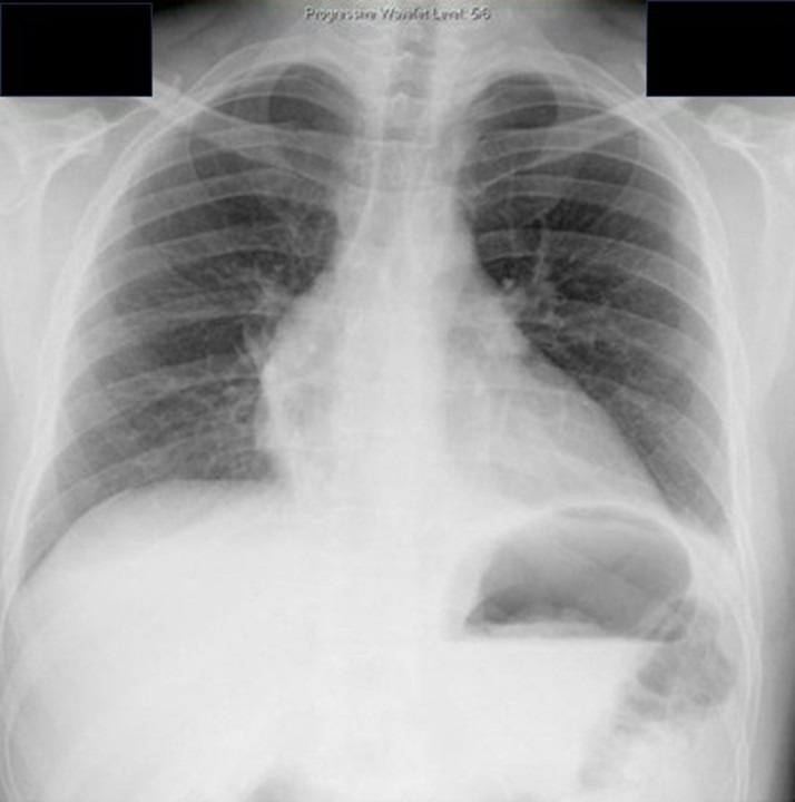
His chest x-ray is shown in Figure 1.


Figure 1. Chest x-ray on the day of injury. To view Figure 1 in a separate enlarged window click here.
Which of the following are true? (Click on the correct answer to be directed to the first of seven pages
Cite as: VonEssen SG. January 2025 Critical Care Case of the Month: A 35-Year-Old Admitted After a Fall. Southwest J Pulm Crit Care Sleep. 2024;30(1):1-4. doi:
October 2024 Critical Care Case of the Month: Respiratory Failure in a Patient with Ulcerative Colitis
Pulmonary Department
Mayo Clinic Arizona
Scottsdale, AZ USA
History of Present Illness
The patient is a 57-year-old woman with a history of ulcerative colitis (UC) complicated by toxic megacolon with subsequent colectomy. She presented to the emergency department with cough, shortness of breath and hypoxemia (87% on RA).
PMH, SH
- UC with history of toxic megacolon (4 years prior) with a total colectomy.
- History of a prior episode of respiratory failure a year earlier thought possibly medication-induced (ustekinumab, Stelara®) which she was taking for her UC. She was treated with steroids with a good response.
- Pyoderma gangrenosum of both ankles (attributed to UC).
- Anemia of chronic disease.
- She is a lifelong non-smoker.
- No exposures to toxic dusts, birds, down, humidifiers, mold or other antigens associated with hypersensitivity pneumonitis.
Physical Exam
- Afebrile, Oxygen saturation 94% on 2 lpm supplemental oxygen.
- Chest: crackles noted at left base.
- CV regular rhythm, no murmur.
- Ext: scarring and erythema on both ankles consistent with resolving pyoderma gangrenosum.
Current Medications
- Clonazepam 1.0 mg daily at bedtime
- Gabapentin 300 mg TID
- Pantoprazole 40 mg BID
- Prednisone 5 mg daily
Laboratory
- Hgb 9.7, WBC 16.9
- Swabs for Influenza A/B and Covid were negative
- Cocci serology negative
A chest radiograph was performed (Figure 1).
 Figure 1. Portable chest X-ray performed in the emergency department. (To view Figure 1 in a separate, enlarged window click here).
Which of the following is/are true regarding the chest X-ray?
- There is a left lower lobe consolidation.
- The portable chest X-ray may be normal.
- A chest CT scan is required to definitely view any consolidation.
- There is a right upper lobe consolidation.
- All of the above.
March 2023 Critical Care Case of the Month: A Bad Egg
Phoenix Pulmonary and Critical Research and Education Foundation
Gilbert, AZ
History of Present Illness
You are asked to see a 35-year-old man who was admitted to the ICU from the ER the previous night with an exacerbation of his chronic obstructive pulmonary disease (COPD). He has a long history of COPD and came to the ER for COVID-19 testing because he was at a party where a friend was later found to COVID-19. He denies any change in his chronic respiratory symptoms but his spirometry was significantly worse than his baseline in the ER and despite his protests he was admitted. He was treated with empiric antibiotics (amoxicillin and clavulanic acid), corticosteroids (methylprednisolone 125 mg every 6 hours), bronchodilators (albuterol/ipratropium every 4 hours) and oxygen. He says his breathing has not improved and he wants to go home. He has had gradually increasing shortness of breath for the past 8-10 years. He has minimal cough but denied any fevers, systemic symptoms, or wheezing.
PMH, FH, and SH
He had a history of multiple pneumothoraces which eventually led to bilateral pleurodesis. He has had not pneumothoraces since. He had a benign bone tumor removed about 25 years ago and a history of manic-depression. There is no FH of any similar type of problems. He does smoke about 3/4 pack of cigarettes per day and has more than occasional marijuana use.
Physical Exam
Physical examination was unremarkable expect for a well-healed scar on the left thigh.
Spirometry
Previous spirometry performed as an outpatient showed his FVC 2.54 L (53% of predicted) with an FEV1 1.25 L (31% of predicted). These improved to 2.99 L and 1.52 L after a bronchodilator. His spirometry last night in the ER was FVC 1.63 L (29 % predicted) and FEV1 0.80 L (18 % predicted).
Radiography
A chest radiograph was performed (Figure 1).
 Figure 1. PA (panel A) and lateral (panel B) chest x-ray.
What should be done at this time? (Click on the correct answer to be directed to the second of five pages)
- Continue his antibiotics, corticosteroids and bronchodilators
- Order an alpha-1 antitrypsin level
- Transfer to the floor
- 1 and 3
- All of the above
April 2018 Critical Care Case of the Month
Clement U. Singarajah, MD
Phoenix VA Medical Center
Phoenix, AZ USA
History of Present Illness
A 70-year-old man was admitted for shortness of breath (SOB) secondary to a “COPD exacerbation/ILD”. A pulmonary consult was placed for possible interstitial lung disease (ILD). A thoracic CT scan for pulmonary embolism showed no embolism and no obvious ILD. He was treated for a COPD exacerbation with the usual therapy of antibiotics, steroids, nebulized bronchodilators and oxygen. He started to improve.
A few days later as he was preparing for discharge, the patient suddenly decompensated becoming more SOB (once more proving that this a dangerous time for patients in hospital). There were reports that this began after he choked and perhaps aspirated on some food and drink. His blood pressure remained stable, but he became tachycardic to 130 beats/min, hypoxic on 100% non-rebreathing mask with saturations of 92%. Obvious clinical acute respiratory failure was present. The patient was started on non-invasive ventilation but continued to deteriorate. He was deemed too unstable to obtain a CT scan. EKG showed sinus tachycardia. The patient was transferred to the ICU for respiratory failure. A chest x-ray was obtained (Figure 1).

Figure 1. Panel A: Admission chest x-ray which was interpreted as not different from the patient’s previous chest x-ray. Panel B: Portable chest x-ray taken shortly after initiation of non-invasive ventilation just after arrival in the intensive care unit.
The portable chest x-ray taken in the ICU shows a new right-sided consolidation and which of the following? (Click on the correct answer to proceed to the second of six pages)
Cite as: Singarajah CU. April 2018 critical care case of the month. Southwest J Pulm Crit Care. 2018;16(4):183-91. doi: https://doi.org/10.13175/swjpcc042-18 PDF
March 2018 Critical Care Case of the Month
Babitha Bijin MD
Jonathan Callaway MD
Janet Campion MD
University of Arizona
Department of Medicine
Tucson, AZ USA
Chief Complaints
- Shortness of breath
- Worsening bilateral LE edema
History of Present Illness
A 53-year-old man with history of multiple myeloma and congestive heart failure presented to the emergency department with complaints of worsening shortness of breath and bilateral lower extremity edema for last 24 hours. In the last week, he has had dyspnea at rest as well as a productive cough with yellow sputum. He describes generalized malaise, loss of appetite, possible fever and notes new bilateral pitting edema below his knees. Per patient, he had flu-like symptoms one week ago and was treated empirically with oseltamivir.
Past Medical History
- Multiple myeloma-IgG kappa with calvarial and humeral metastases, ongoing treatment with cyclophosphamide, bortezomib and dexamethasone
- Community acquired pneumonia 2016, treated with oral antibiotics
- Heart failure with echo 10/2017 showing moderate concentric left ventricular hypertrophy, left ventricular ejection fraction 63%, borderline left atrial and right atrial dilatation, diastolic dysfunction, right ventricular systolic pressure estimated 25 mm Hg
- Hyperlipidemia
- Chronic kidney disease, stage III
Home Medications: Aspirin 81mg daily, atorvastatin 80mg daily, furosemide 10mg daily, calcium / Vitamin D supplement daily, oxycodone 5mg PRN, chemotherapy as above
Allergies: No known drug allergies
Social History:
- Construction worker, not currently working due to recent myeloma diagnosis
- Smoked one pack per day since age 16, recently quit with 30 pack-year history
- Drinks beer socially on weekends
- Married with 3 children
Family History: Mother with hypertension, uncle with multiple myeloma, daughter with rheumatoid arthritis
Review of Systems: Negative except per HPI
Physical Exam
- Vitals: T 39.3º C, BP 80/52, P121, R16, SpO2 93% on 2L
- General: Alert man, mildly dyspneic with speech
- Mouth: Nonicteric, moist oral mucosa, no oral erythema or exudates
- Neck: No cervical neck LAD but JVP to angle of jaw at 45 degrees
- Lungs: Bibasilar crackles with right basilar rhonchi, no wheezing
- Heart: Regular S1 and S2, tachycardic, no appreciable murmur or right ventricular heave
- Abdomen: Soft, normal active bowel sounds, no tendernesses, no hepatosplenomegaly
- Ext: Pitting edema to knees bilaterally, no cyanosis or clubbing, normal muscle bulk
- Neurologic: No focal abnormalities on neurologic exam
Laboratory Evaluation
- Complete blood count: WBC 15.9 (92% neutrophils), Hgb/Hct 8.8/27.1, Platelets 227
- Electrolytes: Na+ 129, K+ 4.0, Cl- 100, CO2 18, blood urea nitrogen 42, creatinine 1.99 (baseline Cr 1.55)
- Liver: AST 35, ALT 46, total bilirubin1.7, alkaline phosphatase 237, total protein 7.4, albumin 2.
- Others: troponin 0.64, brain naturetic peptide 4569, venous lactate 2.6
Chest X-ray

Figure 1. Admission chest x-ray.
Thoracic CT (2 views)

Figure 2. Representative images from the thoracic CT scan in lung windows.
What is most likely etiology of CXR and thoracic CT findings? (Click on the correct answer to proceed to the second of seven pages)
- Coccidioidomycosis pneumonia
- Pulmonary edema
- Pulmonary embolism with infarcts
- Staphylococcus aureus pneumonia
- Streptococcus pneumoniae infection
Cite as: Bijin B, Callaway J, Campion J. March 2018 critical care case of the month. Southwest J Pulm Crit Care. 2018;16(3):117-25. doi: https://doi.org/10.13175/swjpcc035-18 PDF
June 2017 Critical Care Case of the Month
Stephanie Fountain, MD
Pulmonary and Critical Care Medicine
Banner University Medical Center Phoenix
Phoenix, AZ USA
History of Present Illness
The patient is a 60-year-old woman who presented with a month long history of of odynophagia with retrosternal pain and occasional nausea and vomiting.
Past Medical History, Social History and Family History
She has a past medical history of mixed connective tissue disease with anti-phosopholipid antibody. There is also a history of leukocytoclastic vasculitis, chronic leg ulcers, and poor dentition. She also has a history of chronic obstructive lung disease (COPD) and is a current smoker having accumulated about 50 pack-years of cigarette smoking.
Current Medications
- Prednisone 20 mg daily
- Azathioprine 75 mg daily
- Plaquenil 400 mg daily
- Salmeterol/fluticasone BID
- Albuterol prn
Electrocardiographic, Radiologic and Laboratory Evaluation
Her electrocardiogram and chest x-ray were unremarkable. Complete blood count showed a white blood cell count of 10,500 cells per microliter (mcL), hemoglobin 10.3 grams/deciliter (dL), hematocrit 31%, and platelet count of 48,000 cells per mcL. Electrolytes were unremarkable and creatinine was 0.6 mg/dL.
What should be done next? (Click on the correct answer to proceed to the second of six pages)
- Bronchoscopy
- Gastroenterology consult
- Platelet and red blood cell (RBC) transfusion
- 1 and 3
- All of the above
Cite as: Fountain S. June 2017 critical care case of the month. Southwest J Pulm Crit Care. 2017;14(6):262-8. doi: https://doi.org/10.13175/swjpcc061-17 PDF
Tracheal Stoma Necrosis: A Case Report
Stella Pak, MD
Arjan Flora, MD
Young-Sook Yoon, MD
Department of Medicine
University of Toledo Medical Center
Toledo, OH, USA
Abstract
Acute tracheal dilatation, due to an overinflated cuff, has been reported early in the course of mechanical ventilation through an endotracheal tube. Tracheal stoma necrosis is a rare complication, but such can accompany acute tracheal dilation. Herein, we report a case of tracheal necrosis 9 days following tracheostomy placement in a 71-year old woman associated with overinflation of the tracheal tube cuff. This case report aims to 1) add to the scant body of knowledge about the diagnosis and management for the patients with tracheal stoma necrosis and 2) raise awareness for error-traps in interpreting diagnostic images, specifically satisfaction of search error, inattentional blindness error, and alliterative error.
Case Report
A 71-year-old woman with a history of chronic respiratory failure on mechanical ventilation presented to the emergency department for bleeding around the tracheostomy site. The tracheostomy was recently inserted 9 days prior to admission. A chest radiograph demonstrated left lower lobe atelectasis, pleural effusion, and cardiomegaly that was consistent with pre-existing congestive heart failure (Figure 1).

Figure 1. Chest radiograph (AP) performed during first admission.
The cuff overinflation was demonstrated as a spherical shaped hypolucent region surrounding the trachea. However, the lesion escaped attention possibly because the focus of attention was limited to the thoracic compartment. A CT of the soft tissue in the neck ruled out the possibility of hematoma or infection. However, the features suggestive of overinflation of tracheostomy tube once again escaped attention. The spherical shaped hypolucent area, representing the cuff, was 3.9 cm in the anterior-posterior axis and 3.8 cm along the right-left axis.
A fiberoptic bronchoscopy through the tracheostomy tube revealed a large blood clot obstructing the distal end of the tube. A necrotic lesion around the stoma was also found. Careful observation via the bronchoscope during the procedure revealed no tearing or rupture. The patient was conservatively treated with vancomycin and cefepime for treatment of a ventilator-associated pneumonia. The oozing of blood from the tracheostomy stopped on with conservative wound care, including cleaning and dressing. She returned back to her baseline and was subsequently discharged on 3rd day of admission. During this first admission, a tracheostomy tube exchange was not done due to bleeding from the stoma.
The patient was readmitted 12 days after discharge for an episode of hematemesis of approximately 400 mL of bright red blood. A chest radiograph showed satisfactory position of tracheotomy tube and cardiomegaly at baseline (Figure 2).

Figure 2. Chest radiograph (AP) after readmission.
For the third time, the features suggestive of cuff-overinflation went unnoticed, delaying accurate diagnosis and proper treatment.
As a part of the patient’s evaluation, a CT of the chest with intravenous contrast was done, revealing the overinflated cuff of the trachea tube into the soft tissue of the neck (Figure 3).

Figure 3. Thoracic CT scan showing the overinflated tracheostomy cuff in the (A) coronal, (B) sagittal, and (C) axial views.
The ovoid shaped hypolucent area, representing the cuff, was 5.3 cm in the anterior-posterior axis and 4.6 cm along the right-left axis.
The Shiley proximal tracheal tube was urgently replaced with a portex Bivona tracheal tube. The new tracheostomy tube is more extensible, soft, and longer in distal length. Postoperatively, the patient was kept ventilated in the ICU. Repeated chest CT showed the new tracheostomy tube in satisfactory position and normalization of trachea shape. She made an uneventful recovery and was discharged 8 days after the tracheotomy tube replacement.
Discussion
A case of nonfatal hemorrhage due to innominate artery erosion with soft tissue necrosis at the stoma site of a tracheostomy is presented. In this ventilator-dependent patient with a recent tracheotomy stoma creation, an overinflated cuff of a tracheotomy tube was the key culprit in the pathology. Tracheal tube cuff pressure should be monitored so that it does not exceed a reasonable estimate of capillary perfusion pressure. Cuffs with pressure over 25 mmHg can compress the surrounding soft tissue, including delicate vascular structures. The damage to the vasculature in contact with the tube can result in ischemic necrosis in the soft tissue. If left untreated, these necrotic regions can develop infection or undergo fibrosis, leading to progressive stenosis (1).
A number of cognitive errors led to multiple episodes of misdiagnosis in this patient. Satisfaction of search error is a type of false negative error caused by premature termination of search after an abnormality has been detected (2). In this patient, we readily detected several abnormalities—cardiomegaly, pulmonary atelectasis, and pleural effusion. These initial findings likely led us to subconsciously neglect later findings.
Inattentional blindness error is a false negative error caused by the psychological lack of attention on an unexpected stimulus (3). In the present case, none of the diagnostic imaging was taken to check for cuff-overinflation. The images from the first admission were ordered for a concern of NG tube malposition, infection, and hematoma. The images ordered during the second admission were ordered to check the tracheotomy tube position. The thoracic compartment (the area for the expected abnormalities) received a disproportionately large amount of attention, whereas only a scant amount of attention was paid to the neck compartment.
Alliterative error is an error caused by a preconceived notion from a previous interpretation by a colleague or oneself (4). The negative finding in the previous reports could have affected the subsequent interpretative performance.
To the best of our knowledge, there are only 3 other cases of soft tissue necrosis caused by cuff overinflation. In two of these cases, the extended trachea did not recoil back to the previous size (5, 6). In the presented case, the stretched trachea recoiled back, similar to the case described by Sachdeva and his colleagues (7). The prognostic value of this difference in recovery is unknown, but might have a significant clinical implication. To explore the clinical relevance of this finding, more data on this condition is needed.
Teaching Points
- Careful attention should be paid to cuff inflation pressure in patients presenting with bleeding at the tracheostomy site.
- Conscious efforts to avoid well-known errors in diagnostic image interpretation, such as satisfaction of search error, and inattentional blindness error, should be made to improve diagnostic accuracy.
References
- De Leyn P, Bedert L, Delcroix M, et al. Tracheotomy: clinical review and guidelines. Eur J Cardiothorac Surg. 2007 Sep;32(3):412-21. [CrossRef] [PubMed]
- Ashman CJ, Yu JS, Wolfman D. Satisfaction of search in osteoradiology. AJR Am J Roentgenol. 2000 Aug;175(2):541-4. [CrossRef] [PubMed]
- Richards A, Hannon EM, Derakshan N. Predicting and manipulating the incidence of inattentional blindness. Psychol Res. 2010 Nov;74(6):513-23. [CrossRef] [PubMed]
- Berlin L. Malpractice issues in radiology. Alliterative errors. AJR Am J Roentgenol. 2000 Apr;174(4):925-31. [CrossRef] [PubMed]
- Rhodes A, Lamb FJ, Grounds RM, Bennett ED. Tracheal dilatation complicating tracheal intubation. Anaesthesia. 1997 Jan;52(1):70-2. [CrossRef] [PubMed]
- Honig EG, Francis PB. Persistent tracheal dilatation: onset after brief mechanical ventilation with a "soft-cuff" endotracheal tube. South Med J. 1979 Apr;72(4):487-90. [CrossRef] [PubMed]
- Sachdeva A, Pickering EM, Reed RM, Shanholtz CB. Ice cream cone sign: reversible ballooning of the trachea due to tracheostomy tube cuff overinflation. BMJ Case Rep. 2016 May 4;2016. [CrossRef] [PubMed]
Cite as: Pak S, Flora A, Yoon Y-S. Tracheal stoma necrosis: a case report. Southwest J Pulm Crit Care. 2017;14(4):172-6. doi: https://doi.org/10.13175/swjpcc032-17 PDF
Corticosteroids and Influenza A associated Acute Respiratory Distress Syndrome
Philippe R. Bauer, MD, PhD
Vivek N. Iyer, MD, MPH
Pulmonary and Critical Care Medicine
Mayo Clinic
Rochester, MN USA
Abstract
The use of corticosteroids remains controversial in influenza infection, especially with lower respiratory tract infection. We present a case of moderate acute respiratory distress syndrome (ARDS) associated with influenza A that showed a dramatic improvement with combined corticosteroids and antiviral therapy. Host defense against virus infection consists of both innate and adaptive immune responses. An exuberant immune response to the primary pathogen leads to ‘collateral’ lung damage resulting in ARDS. The use of corticosteroids to modulate this excessive immune response, although intuitive, has been associated with increased mortality when administered early in the course of severe influenza A pneumonia. The administration of corticosteroids in this case was associated with a dramatic and unequivocal improvement. This unique case highlights the potential benefits of corticosteroids use in influenza A associated ARDS and may challenge clinicians to rethink current recommendations that specifically discourage corticosteroids use in patients with Influenza A associated ARDS.
Introduction
The impact of corticosteroids on clinical outcome in patients with influenza A associated respiratory failure is unclear (1). Retrospective studies suggest an adverse effect from early parenteral corticosteroids use in patients with pandemic influenza infection. On the other hand, in immunosuppressed patients, high dose corticosteroid given at the time of diagnosis of influenza was associated with a reduced risk for mechanical ventilation, without increased adverse effects other than delayed viral clearance. In general, the effect of corticosteroids on acute respiratory distress syndrome (ARDS) is controversial and its use is not routinely recommended. The adjunctive use of prednisone during the early phase of community-acquired pneumonia may actually reduce the development of ARDS (2). In severe influenza, early corticosteroids showed no evidence of benefit and suggested potential harm (3). We present a case of moderate ARDS associated with influenza A that showed a dramatic and unequivocal improvement after initiation of corticosteroids.
Abbreviations:
APACHE: Acute Physiology and Chronic Health Evaluation
ARDS: Acute Respiratory Distress Syndrome
ICU: Intensive Care Unit
PCR: Polymerase Chain Reaction
SOFA: Sequential Organ Failure Assessment
Case Report
A 62-year old male, nonsmoker, with a history of hypertension, dyslipidemia and depression, presented in March 2014 with chills, fever and nonproductive cough; he was initially treated for ‘bronchitis’ as an outpatient with levofloxacin. He had not received the influenza vaccine. Three days later, he developed acute hypoxemic respiratory failure with bilateral pulmonary infiltrates and was hospitalized elsewhere. Influenza testing was negative and he was started on piperacillin/tazobactam and azithromycin. He was transferred to our facility the next day because of worsening respiratory status. Initial heart rate was 80 bpm, blood pressure was 120/60 mm Hg, respirations was 22/min, and temperature was 37.7 ºC. The Acute Physiology and Chronic Health Evaluation (APACHE) IV score was 55 and the Sequential Organ Failure Assessment (SOFA) score was 8. His presentation was consistent with moderate ARDS with a PaO2/FiO2 ratio of 143, a chest radiograph showing bilateral pulmonary infiltrates (Figure 1) and no evidence of heart failure confirmed by bedside echocardiogram.

Figure 1. Bilateral pulmonary opacities consistent with moderate ARDS (PaO2/FiO2 ratio 143).
Nasal swab was again negative for influenza by polymerase chain reaction (PCR). Leukocyte count was 4.4 x 10⁹/L with lymphopenia (0.22 x 10⁹/L), hemoglobin was 11.7 g/dL, and platelet count was 216 x 109/L. Sodium was 134 mmol/L, creatinine was 1 mg/dL and AST was 142 U/L. He was initiated of high flow nasal oxygen, and vancomycin and oseltamivir were added. Due to the severity of his condition, he was also started on methylprednisolone (125 mg intravenously every 8 hours). After a brief trial of noninvasive ventilation, he was intubated, sedated, paralyzed and placed on a low tidal volume strategy with an initial PEEP of 15 cm H2O and a FiO2 of 0.7. A broncho-alveolar lavage, performed post intubation about 16 hours after admission to our facility, showed 35% alveolar macrophages, 8% lymphocytes and 57% neutrophils and was positive for influenza A by PCR; cultures were negative for other organisms. Other tests including HIV, RSV, Mycoplasma, Legionella and urine for Streptococcus antigen were all negative. The patient improved rapidly. He was extubated two days later, and continued on prednisone (40 mg daily) for five more days when he was dismissed home without any need for supplemental oxygen, although the chest radiograph continued to show infiltrates.
Discussion
This case illustrates a patient with delayed diagnosis and treatment of influenza A associated with moderate ARDS who made a rapid and complete recovery with antiviral, antibiotic and adjunctive high dose corticosteroid therapy.
The diagnosis of influenza A in this case meets all criteria established by Clinical Practice Guidelines of the Infectious Diseases Society of America (4). Rapid influenza testing lack sensitivity and false negative are not infrequent. ARDS is a well-defined complication of influenza infection. While the administration of corticosteroids appeared to temporally co-relate with clinical improvement, a causal link cannot be established definitively. The role of immunosuppression in influenza associated ARDS is very controversial with conflicting evidence from prospective (supportive) and retrospective (against) studies. For example, the combined use of sirolimus and prednisone was associated with significantly improved oxygenation as well as reduced organ dysfunction in mechanically ventilated patients with severe H1N1 respiratory failure (5). On the other hand, retrospective studies have shown increased mortality with the early use of high dose corticosteroids in severe influenza A pneumonia and respiratory failure. Furthermore, corticosteroids are now rarely used in ARDS and only sparingly given in case of refractory septic shock. The immune response to influenza infection depends on the virus, the host and the host response to infection. Host defense against virus infection consists of both innate and adaptive immune responses. An excessive immune response may result in ‘collateral damage’ and critical respiratory illness which may be ameliorated by the use of systemic corticosteroids. On the other hand, suppression of the host immune system may enhance viral replication and prolong critical illness. As a result of these conflicting data, major societies have been unable to firmly recommend for or against corticosteroids therapy in Influenza A associated respiratory failure.
In conclusion, we report on a case of Influenza A with ARDS and rapid improvement on corticosteroids. We have reviewed the current uncertainty surrounding the use of corticosteroids in this setting and leave open the possibility for careful consideration of this adjunctive therapy in other cases. Randomized trials are needed to further delineate the potential benefit of corticosteroids in severe influenza infection.
References
- Rodrigo C, Leonardi-Bee J, Nguyen-Van-Tam J, Lim WS. Corticosteroids as adjunctive therapy in the treatment of influenza. Cochrane Database Syst Rev. 2016 Mar 7;3:CD010406. [CrossRef] [PubMed]
- Blum CA, Nigro N, Briel M, et al. Adjunct prednisone therapy for patients with community-acquired pneumonia: a multicentre, double-blind, randomised, placebo-controlled trial. Lancet. 2015 Apr 18;385(9977):1511-8. [CrossRef] [PubMed]
- Brun-Buisson C, Richard JC, Mercat A, Thiébaut AC, Brochard L; REVA-SRLF A/H1N1v 2009 Registry Group. Early corticosteroids in severe influenza A/H1N1 pneumonia and acute respiratory distress syndrome. Am J Respir Crit Care Med. 2011 May 1;183(9):1200-6. [CrossRef] [PubMed]
- Harper SA, Bradley JS, Englund JA, et al. Seasonal influenza in adults and children--diagnosis, treatment, chemoprophylaxis, and institutional outbreak management: clinical practice guidelines of the Insert LinkInfectious Diseases Society of America. Clin Infect Dis. 2009 Apr 15;48(8):1003-32. [CrossRef] [PubMed]
- Wang CH, Chung FT, Lin SM, Huang SY, Chou CL, Lee KY, Lin TY, Kuo HP. Adjuvant treatment with a mammalian target of rapamycin inhibitor, sirolimus, and steroids improves outcomes in patients with severe H1N1 pneumonia and acute respiratory failure. Crit Care Med. 2014 Feb;42(2):313-21. [CrossRef] [PubMed]
Cite as: Bauer PR, Iyer VN. Corticosteroids and influenza A associated acute respiratory distress syndrome. Southwest J Pulm Crit Care. 2016;13(5):248-51. doi: https://doi.org/10.13175/swjpcc102-16 PDF
October 2016 Critical Care Case of the Month
Stephanie Fountain, MD
Banner University Medical Center Phoenix
Phoenix, AZ USA
Critical Care Case of the Month CME Information
Members of the Arizona, New Mexico, Colorado and California Thoracic Societies and the Mayo Clinic are able to receive 0.25 AMA PRA Category 1 Credits™ for each case they complete. Completion of an evaluation form is required to receive credit and a link is provided on the last panel of the activity.
0.25 AMA PRA Category 1 Credit(s)™
Estimated time to complete this activity: 0.25 hours
Lead Author(s): Stephanie Fountain, MD. All Faculty, CME Planning Committee Members, and the CME Office Reviewers have disclosed that they do not have any relevant financial relationships with commercial interests that would constitute a conflict of interest concerning this CME activity.
Learning Objectives:
As a result of this activity I will be better able to:
- Correctly interpret and identify clinical practices supported by the highest quality available evidence.
- Will be better able to establsh the optimal evaluation leading to a correct diagnosis for patients with pulmonary, critical care and sleep disorders.
- Will improve the translation of the most current clinical information into the delivery of high quality care for patients.
- Will integrate new treatment options in discussing available treatment alternatives for patients with pulmonary, critical care and sleep related disorders.
Learning Format: Case-based, interactive online course, including mandatory assessment questions (number of questions varies by case). Please also read the Technical Requirements.
CME Sponsor: University of Arizona College of Medicine
Current Approval Period: January 1, 2015-December 31, 2016
Financial Support Received: None
A 27-year-old Caucasian man with past medical history of opioid abuse (reportedly sober for 10 years on buprenorphine), post traumatic stress disorder, depression and anxiety presented to the emergency department complaining of dysarthria after taking diphenhydramine and meclizine in addition to his prescribed trazodone and buprenorphine to try to sleep. He was discharged to home after his symptoms appeared to improve with intravenous fluid.
He returned to the emergency department the following afternoon with worsening dysarthria, dysphagia, and subjective weakness. The patient was non toxic appearing, afebrile, vital signs were stable and his strength was reported as 5/5. Computed tomography of his head did not show any evidence of acute intracranial abnormality. Given his ongoing complaints, he was admitted for observation to the general medicine wards.
That night a rapid response was initiated when the nurse found the patient to be unresponsive, but spontaneously breathing. The patient’s clinical status did not change with naloxone administration. An arterial blood gas obtained demonstrated a profound respiratory acidosis with a pH of 7.02 and a pCO2 of 92. He was emergently intubated. A chest x-ray was performed (Figure 1).

 Figure 1. Panel A: admission portable chest x-ray. Panel B: chest -ray immediately after intubation.
Which of the following are present on his chest X-ray? (Click on the correct answer to proceed to the second or four panels)
Cite as: Fountain S. October 2016 critical care case of the month. Soutwest J Pulm Crit Care. 2016:13(4):159-64. doi: http://dx.doi.org/10.13175/swjpcc095-16 PDF
September 2016 Critical Care Case of the Month
Clement U. Singarajah, MD
Samir Sultan, DO
Phoenix VA Medical Center
Phoenix, AZ USA
Critical Care Case of the Month CME Information
Members of the Arizona, New Mexico, Colorado and California Thoracic Societies and the Mayo Clinic are able to receive 0.25 AMA PRA Category 1 Credits™ for each case they complete. Completion of an evaluation form is required to receive credit and a link is provided on the last panel of the activity.
0.25 AMA PRA Category 1 Credit(s)™
Estimated time to complete this activity: 0.25 hours
Lead Author(s): Clement U. Singarajah, MD. All Faculty, CME Planning Committee Members, and the CME Office Reviewers have disclosed that they do not have any relevant financial relationships with commercial interests that would constitute a conflict of interest concerning this CME activity.
Learning Objectives:
As a result of this activity I will be better able to:
- Correctly interpret and identify clinical practices supported by the highest quality available evidence.
- Will be better able to establsh the optimal evaluation leading to a correct diagnosis for patients with pulmonary, critical care and sleep disorders.
- Will improve the translation of the most current clinical information into the delivery of high quality care for patients.
- Will integrate new treatment options in discussing available treatment alternatives for patients with pulmonary, critical care and sleep related disorders.
Learning Format: Case-based, interactive online course, including mandatory assessment questions (number of questions varies by case). Please also read the Technical Requirements.
CME Sponsor: University of Arizona College of Medicine
Current Approval Period: January 1, 2015-December 31, 2016
Financial Support Received: None
Clinical History
A 66-year-old man was admitted to the ICU in complete heart block with borderline hypotension. After cardiology consultation, a decision was made to place an urgent transvenous pacer. The transvenous pacer was place without use fluoroscopy from an right internal jugular venous (IJV) approach using real time ultrasound by two very experienced operators. The ultrasound confirmed right IJV placement and the pacer was found to capture and pace appropriately without any complications. A post placement CXR was obtained (Figure 1).

Figure 1. Portable chest x-ray after RIJV transvenous pacer (TVP).
What does the chest x-ray show? (Click on the correct answer to proceed to the second of five panels)
Cite as: Singarjah CU, Sultan S. September 2016 critical care case of the month. Southwest J Pulm Crit Care. 2016;13(3):108-13. doi: http://dx.doi.org/10.13175/swjpcc079-16 PDF
March 2016 Critical Care Case of the Month
Theo Loftsgard APRN, ACNP
Joel Hammill APRN, CNP
Mayo Clinic Minnesota
Rochester, MN USA
Critical Care Case of the Month CME Information
Members of the Arizona, New Mexico, Colorado and California Thoracic Societies and the Mayo Clinic are able to receive 0.25 AMA PRA Category 1 Credits™ for each case they complete. Completion of an evaluation form is required to receive credit and a link is provided on the last panel of the activity.
0.25 AMA PRA Category 1 Credit(s)™
Estimated time to complete this activity: 0.25 hours
Lead Author(s): Theo Loftsgard APRN, ACNP. All Faculty, CME Planning Committee Members, and the CME Office Reviewers have disclosed that they do not have any relevant financial relationships with commercial interests that would constitute a conflict of interest concerning this CME activity.
Learning Objectives:
As a result of this activity I will be better able to:
- Correctly interpret and identify clinical practices supported by the highest quality available evidence.
- Will be better able to establsh the optimal evaluation leading to a correct diagnosis for patients with pulmonary, critical care and sleep disorders.
- Will improve the translation of the most current clinical information into the delivery of high quality care for patients.
- Will integrate new treatment options in discussing available treatment alternatives for patients with pulmonary, critical care and sleep related disorders.
Learning Format: Case-based, interactive online course, including mandatory assessment questions (number of questions varies by case). Please also read the Technical Requirements.
CME Sponsor: University of Arizona College of Medicine
Current Approval Period: January 1, 2015-December 31, 2016
Financial Support Received: None
History of Present Illness
A 58-year-old man was admitted to the ICU in stable condition after an aortic valve replacement with a mechanical valve.
Past Medical History
He had with past medical history significant for endocarditis, severe aortic regurgitation related to aortic valve perforation, mild to moderate mitral valve regurgitation, atrial fibrillation, depression, hypertension, hyperlipidemia, obesity, and previous cervical spine surgery. As part of his preop workup, he had a cardiac catheterization performed which showed no significant coronary artery disease. Pulmonary function tests showed an FEV1 of 55% predicted and a FEV1/FVC ratio of 65% consistent with moderate obstruction.
Medications
Amiodarone 400 mg bid, digoxin 250 mcg, furosemide 20 mg IV bid, metoprolol 12.5 mg bid. Heparin nomogram since arrival in the ICU.
Physical Examination
He was extubated shortly after arrival in the ICU. Vitals signs were stable. His weight had increased 3 Kg compared to admission. He was awake and alert. Cardiac rhythm was irregular. Lungs had decreased breath sounds. Abdomen was unremarkable.
Laboratory
His admission laboratory is unremarkable and include a creatinine of 1.0 mg/dL, blood urea nitrogen (BUN) of 18 mg/dL, white blood count (WBC) of 7.3 X 10⁹ cells/L, and electrolytes with normal limits.
Radiography
His portable chest x-ray is shown in Figure 1.

Figure 1. Portable chest x-ray taken on admission to the ICU.
What should be done next? (Click on the correct answer to proceed to the second of five panels)
- Bedside echocardiogram
- Diuresis with a furosemide drip because of his weight gain and cardiomegaly
- Observation
- 1 and 3
- All of the above
Cite as: Loftsgard T, Hammill J. March 2016 critical care case of the month. Southwest J Pulm Crit Care. 2016;12(3):81-8. doi: http://dx.doi.org/10.13175/swjpcc018-16 PDF
January 2016 Critical Care Case of the Month
Sandra L. Till, DO
Banner University Medical Center Phoenix
Phoenix, AZ USA
History of Present Illness
The patient is an 18-year-old woman who was driving to high school on a frontage road when she fell asleep at the wheel and her car rolled over. She was wearing her seatbelt but there was no airbag deployment. She did not lose consciousness and she was responsive and answering questions at the scene. She self-extricated from the vehicle. She had left arm pain with a boney deformity and she walked to the ambulance that transferred her to the hospital emergency department (ED).
Upon arrival in the ED she appeared pale and had difficulty breathing. In addition to her arm pain with an obvious left humeral fracture she also complained of upper abdominal and anterior chest pain. O2 saturation was initially 90% but declined to 70%.
Which of the following should be ordered immediately? (Click on the correct answer to proceed to the second of six panels)
- Begin intravenous lines with large bore needles
- X-ray of humerus
- Hemoglobin and hematocrit
- 1 and 3
- All of the above
Cite as: Till SL. January 2016 critical care case of the month. Southwest J Pulm Crit Care. 2016;12:6-12. doi: http://dx.doi.org/10.13175/swjpcc151-15 PDF
September 2015 Critical Care Case of the Month: If You Don't Look, You Won't Find
Robert A. Raschke, MD
Banner University Medical Center
Phoenix, AZ
History of Present Illness
A 55-year-old woman was transferred from Mexico emergently for acute cardiomyopathy. On the day of admission, she went for a 45-min “exercise” walk and cleaned her house. While taking a shower, she suffered an acute onset of dyspnea with nausea and vomiting and possibly a small amount of hematemesis. She appeared seriously ill to her husband, who took her blood pressure (198/?) and pulse (90) and rushed her to a local medical facility. There, she was found to have severe pulmonary edema, and a troponin of 11. Her echo showed inferior wall motion abnormality with an ejection fraction of 35%. However, coronary catheterization showed normal coronaries. She was treated with oxygen, furosemide, labetolol and enoxaparin and transferred emergently to Banner-University Medical Center.
Past Medical History, Family History and Social History
The patient reported intermittent "spells" since May. These typically occurred upon lying down in bed and were characterized by her as a feeling of “numbness” or tingling which ascends from her chest to her head associated with palpitations and a feeling of “desperation”, typically relieved after a few minutes upon getting up out of bed.
She had a history of hypertension and had been on losartan but this was discontinued a few months previously because of the onset of orthostatic dizziness. She also has a history of hypothyroidism and is taking synthroid. She was treated three times in the last 6 month for amoebiasis. She is a medical missionary to La Paz, Mexico and has recently traveled to Bolivia and Guatemala.
Review of Systems
She has had some night sweats, coughing with deep inspiration, and some slight hemoptysis. She did have a headache one month previously at 7000 ft elevation while in Guatemala.
Physical Examination
- She appears in moderate distress. Her vital signs are normal other than a mild tachycardia.
- She does have rales on auscultation of her lungs.
- The remainder of the physical examination was unremarkable.
Radiography
A portable chest radiograph is performed (Figure 1).

Figure 1. Admission portable chest radiograph.
Laboratory evaluation
Her CBC shows a normal hemoglobin and hematocrit but with an elevated white blood cell count of 26,500 cells/mcL with a left shift. Admission electrolytes and blood sugar are within normal limits.
What additional procedures/testing are indicated? (Click on the correct answer to proceed to the second of four panels)
- Blood cultures
- Echocardiogram
- Electrocardiogram
- NT-pro-brain natriuretic peptide (NT-pro-BNP)
- All of the above
Reference as: Raschke RA. September 2015 critical care case of the month: if you don't look, you won't find. Southwest J Pulm Crit Care. 2015;11(3):97-102. doi: http://dx.doi.org/10.13175/swjpcc113-15 PDF
August 2015 Critical Care Case of the Month: A Diagnostic Branch of Medicine
William T. Love, MD
Karen L. Swanson, DO
Department of Pulmonary Medicine
Mayo Clinic Arizona
Scottsdale, AZ
History of Present Illness
A 66-year-old man had undergone an orthotopic heart transplantation on March 28th, 2015 due to end-stage cardiomyopathy. During a recent hospitalization from 6/26-7/2 a transbronchial lung biopsy was suggestive of subacute rejection. He was treated with:
- Plasmapheresis x 3
- Intravenous immunoglobulin (IVIG)
- 500 mg Solu-Medrol daily
- Tacrolimus held as supra-therapeutic level of 16.2
- Mycophenolate decreased to 500mg BID
- Prednisone at 10mg BID on discharge
On July 3rd he began having cough productive of clear sputum, nausea, vomiting, and headache. Subsequently he had body aches, subjective fever, chills, night sweats, and a poor appetite with a 4 kg weight loss over the last week. There was also a history of several falls after “losing his balance".
Past Medical History
There was also a history of type 2 diabetes mellitus, chronic kidney disease, coronary artery disease with coronary artery bypass grafting in 2000.
Physical Examination
- Vital signs: T-37.1, HR-100, BP-130/88, RR-22, 96% RA
- Heart: regular rate & rhythm. 2/6 Systolic Murmur
- Lungs: clear to auscultation bilaterally
Laboratory
- Hemoglobin 9.7, WBC 6.3, creatinine 2.2, mildly elevated AST/ALT
- Lumbar Puncture– Protein 58 mg/dL, Glucose 46 mg/dL, 47 Nucleated cells
Radiography
A chest x-ray was performed (Figure 1).

Figure 1. Admission PA of the chest.
Based on the chest x-ray and lumbar puncture, which of the following are true? (Click on the correct answer to proceed to the second of four panels)
- The chest x-ray and lumbar puncture findings in this clinical situation suggest cancer metastatic to the lung and brain
- The chest x-ray and lumbar puncture findings in this clinical setting suggest an infection involving the lung and brain
- The clinical findings suggest granulomatosis with polyangiitis (formerly known as Wegener's granulomatosis)
- The clinical findings are suggestive of acute rejection
- The clinical findings are suggestive of tuberculosis
Reference as: Love WT, Swanson KL. August 2015 critical care case of the month: a diagnostic branch of medicine. Southwest J Pulm Crit Care. 2015;11(2):59-65. doi: http://dx.doi.org/10.13175/swjpcc100-15 PDF
June 2015 Critical Care Case of the Month: Just Ask the Nurse
Robert A. Raschke, MD
Banner University Medical Center
Phoenix, AZ
History of Present Illness
A 61-year-old police officer had just finished delivering a speech at a law enforcement conference in Phoenix when he briefly complained of chest pain or chest tingling before lapsing into a mute state. He became diaphoretic cyanotic, and vomited. Emergency medical services was called. They noted a blood pressure of 80/50 mm Hg, a pulse of 45, temperature of 95º F, a respiratory rate of 12, and widely dilated pupils. He was transported to the emergency room.
PMH, SH, FH, Medications
Unknown.
Physical Examination
Vital signs: blood pressure 120/75 mm Hg by oscillometric thigh cuff, pulse 43 and irregular, temperature 96º F, respiratory rate 10, SpO2 96% on O2 @ 5L/min by nasal cannula
Neck: No JVD.
Lungs: Poor inspiratory effort
Heart: Irregular rhythm without a murmur
Neurological:
- Delirious – mute – won’t obey commands or track with his eyes
- Pupils 3 mm reactive
- Withdrew 3 extremities to nail bed pressure – he will defend his left arm with his right arm
He suddenly became asystolic and cardiopulmonary resuscitation was begun. After about a minute a femoral pulse could be felt.
Which of the following are indicated at this time? (Click on the correct answer to proceed to the second of five panels)
Reference as: Raschke RA. June 2015 critical care case of the month: just ask the nurse. Southwest J Pulm Crit Care. 2015;10(6):323-9. doi: http://dx.doi.org/10.13175/swjpcc077-15 PDF
Analysis of a Fatal Left Ventricular Assist Device Infection: A Case Report and Discussion
Neal Stuart Gerstein, MD FASE1
Henry G. Chou, MD2
Andrew Lewis Dixon, MD1
1Department of Anesthesiology & Critical Care Medicine
University of New Mexico
Albuquerque, NM
2Department of Anesthesiology
Cedars-Sinai Medical Center
Los Angeles, CA
Introduction
Left ventricular assist device (VAD) therapy is an increasingly utilized treatment as a bridge to heart transplantation or as long-term destination therapy. Recent reports show there is a 22% - 32% incidence of VAD-associated infections with staphylococci and nosocomial gram-negative bacilli being the most common causative organisms (1,2). These organisms are often found in intensive care units, where they have the highest proportion of resistance, thus exposing already critically ill patients to the possibility of resistant organism VAD-associated infections (3). Mortality rates exceed 60% when sepsis develops in a patient with a continuous flow left VAD and infection is the number one cause of death in those awaiting cardiac transplantation (4,5). With continued left VAD use clinicians will likely see multidrug-resistant (MDR) or even pandrug-resistant organism VAD-associated infections. Clinicians need to be prepared to manage such an intimidating entity.
Case Report
We report a case of a 25 year-old male with a pandrug-resistant Pseudomonas aeruginosa VAD-associated infection. The patient’s medical history is significant for a diagnosis of idiopathic dilated cardiomyopathy refractory to maximal medical therapy requiring implantation of a HeartMate II (Thoratec Co., Pleasanton, CA, USA) continuous flow left VAD (Figure 1).

Figure 1. HeartMate II® left VAD schematic (reprinted with the permission of Thoratec Co., Pleasanton, CA, USA).
His course was complicated with multiple hospital admissions for recurrent VAD-associated infections and numerous episodes of P. aeruginosa bacteremia that had been treated with a multitude of antipseudomonal antibiotics. He presented to our hospital for management of severe volume overload in the setting of VAD-associated infections. Transesophageal echocardiography demonstrated a left ventricular ejection fraction of 24% with severe left and right ventricular dilatation. Chest x-ray revealed cardiomegaly and multiple devices including the left VAD (Figure 2).

Figure 2. Chest X-ray demonstrating an enlarged cardiac silhouette, the HeartMate II axial pump (*) with inflow (down arrow, ↓) and outflow (up arrow, ↑) cannulas, biventricular pacer with leads in right atrium (A), coronary sinus (B), and right ventricle (C) (dashed arrows).
Blood cultures revealed MDR P. aeruginosa; except for showing intermediate sensitivity to tobramycin there was resistance to all antimicrobials tested. In vitro synergy testing revealed modest bacterial inhibition when only colistin, fosfomycin, imipenem, and tobramycin were combined. After maximizing medical therapy, multiple left VAD pocket washings and implantation of tobramycin beads followed. Intraoperative findings included an encapsulated infection around the driveline and obvious infection of the left VAD pocket. Repeat blood cultures showed P. aeruginosa had developed resistance to all antimicrobials including tobramycin. Subsequently the left VAD was explanted and the patient was transitioned to an extracorporeal membrane oxygenator (ECMO) in attempt to clear the infection. He was then transitioned to a TandemHeart (CardiacAssist Inc., Pittsburgh, PA, USA), a percutaneous LVAD, as he was not dependent on ECMO for oxygenation. He was able to clear the bacteremia after removal of the infected HeartMate II while on colistin, fosfomycin, tobramycin, azithromycin and rifampin, but was not able to clear the remaining left VAD pocket infection, which again spread systemically. Despite maximal medical and surgical interventions, he died from profound septic shock and multisystem organ failure. To date this is the first known case of a pandrug-resistant P. aeruginosa VAD-associated infection reported in the literature.
Discussion
P. aeruginosa organisms have intrinsic resistance to numerous broad spectrum antibiotics, and can easily develop acquired resistance to most if not all available antimicrobial agents (3). Risk factors for the development of pandrug-resistant P. aeruginosa include previous treatment with antipseudomonal antibiotics and prolonged treatment times. Given our patient had multiple P. aeruginosa infections, treated with multiple rounds of antipseudomonal antibiotics, it is not surprising that pandrug-resistance developed. Few therapeutic options are available for treatment and no new agents are available to evade the known resistance mechanisms. Treatment can be optimized using synergistic combination therapy, which may be the only medical management option in patients with pandrug-resistant P. aeruginosa infections. Some have suggested that rifampin in combination with colistin may be a promising approach (3). Some experts recommend in vitro synergy testing when an organism is resistant to currently recommended antibiotic regimens (6,7). However, a recent review of antibiotic therapy for gram-negative infections describes the utility of in vitro synergy testing equivocal in the context of Pseudomonas infection (8). We managed our patient with combination therapy; however, not until pandrug-resistant P. aeruginosa was isolated did we introduce rifampin in combination with colistin.
A recent review of VAD-associated infections showed the majority were managed without surgical intervention; only 13% required surgical debridement and only in cases of severe infection and/or failed conservative treatment was left VAD explantation required. Since this case there has been a proposed algorithm for management of VAD-associated infections (2); our management, though prior to published guidelines, was in step with the algorithm. Of note, there was no discussion of explanting an left VAD to ECMO to aid in clearing a resistant infection. We felt this was a rational option given our inability to clear the infection. It is unclear as to exactly why our patient was never able to fully clear his infection. Given the patient’s other pre-existing extensive cardiac hardware (i.e. implanted pacer), it is possible that he remained colonized even after maximal surgical and medical therapy. Though speculative, it is possible that removing all foreign material may have allowed for complete infection clearance.
Aside from aggressive medical and surgical management, systolic heart failure with VAD-associated infections may be effectively managed with heart transplantation (9). Our consensus was that this option was neither in the best interest of the patient nor the best use of available resources given the severity of his condition.
Conclusion
Clinicians will continue to see VAD-associated infections with resistant organisms. To minimize adverse outcomes, including VAD-associated infection, prudent patient selection and timing of VAD placement is paramount, as VAD’s placed in critically ill patients have been consistently associated with adverse outcomes (10).
References
- Gordon RJ, Weinberg AD, Pagani FD, Slaughter MS, Pappas PS, Naka Y, Goldstein DJ, Dembitsky WP, Giacalone JC, Ferrante J, Ascheim DD, Moskowitz AJ, Rose EA, Gelijns AC, Lowy FD. Prospective, multicenter study of ventricular assist device infections. Circulation. 2013;127:691-702. [CrossRef] [PubMed]
- Nienaber JJ, Kusne S, Riaz T, Walker RC, Baddour LM, Wright AJ, Park SJ, Vikram HR, Keating MR, Arabia FA, Lahr BD, Sohail MR. Clinical manifestations and management of left ventricular assist device-associated infections. Clin Infect Dis. 2013;57:1438-48. [CrossRef] [PubMed]
- Zavascki AP, Carvalhaes CG, Picao RC, Gales AC. Multidrug-resistant Pseudomonas aeruginosa and Acinetobacter baumannii: resistance mechanisms and implications for therapy. Expert Rev Anti Infect Ther. 2010;8:71-93. [CrossRef] [PubMed]
- Topkara VK, Kondareddy S, Malik F, Wang IW, Mann DL, Ewald GA, Moazami N. Infectious complications in patients with left ventricular assist device: etiology and outcomes in the continuous-flow era. Ann Thorac Surg. 2010;90:1270-7. [CrossRef] [PubMed]
- Bartoli CR, Demarest CT, Khalpey Z, Takayama H, Naka Y. Current management of left ventricular assist device erosion. J Card Surg. 2013;28:776-82. [CrossRef] [PubMed]
- Balaji V, Jeremiah SS, Baliga PR. Polymyxins: Antimicrobial susceptibility concerns and therapeutic options. Indian J Med Microbiol. 2011;29:230-42. [CrossRef] [PubMed]
- Martis N, Leroy S, Blanc V. Colistin in multi-drug resistant Pseudomonas aeruginosa blood-stream infections: a narrative review for the clinician. J Infect. 2014;69:1-12. [CrossRef] [PubMed]
- Tamma PD, Cosgrove SE, Maragakis LL. Combination therapy for treatment of infections with gram-negative bacteria. Clin Microbiol Rev. 2012;25:450-70. [CrossRef] [PubMed]
- Prendergast TW, Todd BA, Beyer AJ, 3rd, Furukawa S, Eisen HJ, Addonizio VP, Browne BJ, Jeevanandam V. Management of left ventricular assist device infection with heart transplantation. Ann Thorac Surg. 1997;64:142-7. [CrossRef] [PubMed]
- Lietz K, Long JW, Kfoury AG, Slaughter MS, Silver MA, Milano CA, Rogers JG, Naka Y, Mancini D, Miller LW. Outcomes of left ventricular assist device implantation as destination therapy in the post-REMATCH era: implications for patient selection. Circulation. 2007;116:497-505. [CrossRef] [PubMed]
Reference as: Gerstein NS, Chou HG, Dixon AL. Analysis of a fatal left ventricular assist device infection: a case report and discussion. Southwest J Pulm Crit Care. 2015;10:16-20. doi: http://dx.doi.org/10.13175/swjpcc139-14 PDF
January 2015 Critical Care Case of the Month: Who's Your Momma?
Carlos Hartmann, MD
Layth Al-Jashaami, MD
Timothy T. Kuberski, MD
Department of Medicine
Maricopa Integrated Health Services
Phoenix, AZ USA
History of Present Illness
A 39-year-old Hispanic woman was admitted complaining of shortness of breath and bilateral lower extremity edema. She was felt to be in acute hypoxic respiratory failure.
Past Medical History
- Systemic lupus erythematosus
- Congestive heart failure with an ejection fraction of 40%
- End-stage renal disease on dialysis secondary to lupus nephritis
Medications
- Rituximab 550 mg once a week
- Prednisone 40 mg daily
- Plaquenil 200 mg twice a day
Physical Examination
The patient was tachypneic in obvious respiratory distress. She was afebrile. Crackles at the bases. Heart was tachycardic. There was 3+ bilateral pitting pretibial edema.
Laboratory
CBC: Hemoglobin 7.8 g/dL, WBC 11 X 109 cells per liter, differential: neutrophils 98%, eosinophils 0%, lymphocytes 1%.
Electrolytes: Potassium 5.8 mEq/L, sodium 143 mEq/L, creatinine 3.3 g/dL, BUN 98 mg/dL.
Brain naturetic peptide: 4055 pg/ml.
Imaging
Admission chest x-ray showed cardiomegaly and bilateral interstitial prominence suggestive of congestive heart failure.
Which of the following are appropriate initial management? (Click on the correct answer to proceed to the 2nd of 4 panels)
- Bronchoscopy with bronchoalveolar lavage
- Hemodialysis
- Increased methylprednisolone for a potential lupus "flare"
- 1 and 3
- All of the above
Reference as: Hartmann C, Al-Jashaami L, Kuberski TT. January 2015 critical care case of the month: who's your momma? Southwest J Pulm Crit Care. 2015:10(1):11-15. doi: http://dx.doi.org/10.13175/swjpcc145-14 PDF
June 2014 Critical Care Case of the Month: Acute Exacerbation in Cystic Fibrosis
Seongseok Yun, MD PhD1
Juhyung Sun, BS2
Laura Howe, MD1
Roberto Bernardo, MD1
Sepehr Daheshpour, MD1
Department of Medicine1
College of Medicine2
University of Arizona
Tucson, AZ 85724
History of Present Illness
A 28 year-old woman with a history of cystic fibrosis, presented with worsening shortness of breath and cough associated with productive secretions. She was diagnosed with cystic fibrosis when she was 14 months old, and has a history of multiple inpatient admissions for acute pulmonary exacerbation of cystic fibrosis. Her most recent hospitalization was a month prior to this admission, and sputum culture demonstrated methicillin-resistant Staphylococcus aureus, multidrug-resistant Pseudomonas aeruginosa, and Achromobacter xylosoxidans. She was treated with linezolide, meropenum, colistin, and azithromycin with significant symptom improvement, then, discharged home with ciprofloxacin, linezolide and zosyn. However, she developed worsening respiratory distress again and came back to hospital. In the emergency department she required 10 L/min of oxygen to maintain an SpO2 above 90 %.
PMH
- Cystic fibrosis
- Seizure
- Kidney stone
- Portacath placement
- Gastrostomy tube placement
Medications
- Azithromycin 500 mg 3 times a day
- Dornase alpha 1 mg/ml nebulizer twice a day
- Fluticasone-salmeterol 500-50 mcg/dose inhaler twice a day
- Lipase-protease-amylase 21,000-37,000-61,000 unit 4 caps a day
- Cholecalciferol 2,000 unit capsule daily
- Ferrous sulfate 325 mg PO twice a day
- Ascorbic acid 250 mg PO twice a day
- Oxycodone-acetaminophen 10-325 mg 4 times a day as needed
Social History
- No smoking
- No alcohol use
- No recreational drug use
Physical Examination
Vital signs: Temperature 37.3 °C, heart rate 114 beats/min, respiratory rate 20-24 breaths/min, blood pressure 99/69mmHg, SpO2 88-90 % on 10 L NC
General: Alert and oriented X 3, acutely distressed, tachypneic and dyspneic
Skin: Diaphoretic. No rash or lesions.
HEENT: Unremarkable.
Respiratory: Diffuse rales in all lung fields, no wheezing, no stridor
CVS: Tachycardic, regular rhythm, no murmur.
Abdomen: Soft, non-tender, no tenderness, no guarding, no hepato-splenomegaly, PEG tube placed
Lymphatics: No cervical or axillary lymphadenopathy
Extremities: No clubbing, no cyanosis, no peripheral edema, normal tone, normal range of movement
Neurological: Normal speech, no focal neurologic deficit, CN exam within normal range
Laboratory
CBC: WBC 11.9X 10³ /μL, Hb 9.8 g/dL, Hct 30.7%, Platelets 356,000 /μL.
Chemistries: Na+ 137 meq/L, K+ 4.1 meq/L, Cl- 107 meq/L, CO2 22 mmol/L, blood urea nitrogen (BUN) 13 mg/dL, creatinine 0.7 mg/dL, glucose 106 mg/dL, calcium 8.0 mg/dL, albumin 2.6 g/dL, liver function tests within normal limits.
Prothrombin time (PT) 14.0 sec, international normalized ratio (INR)1.1, partial thromboplastin time (PTT) 37.2sec
Pulmonary Function Test
FVC 48 % (1.95 L), FEV1 36 % (1.25 L), FEF25-75 14 % (0.55 L/sec)
Radiography
An old chest x-ray and thoracic CT scan were reviewed (Figure 1).

Figure 1. Previous PA (Panel A), lateral (Panel B) chest x-ray and representative image from the thoracic CT scan (Panel C).
Which of the following are findings of cystic fibrosis on chest x-ray? (Click on the correct answer to move to the next panel)
Reference as: Yun S, Sun J, Howe L, Bernardo R, Daheshpour S. June 2014 critical care case of the month: acute exacerbation in cystic fibrosis. Southwest J Pulm Crit Care. 2014;8(6):305-19. doi: http://dx.doi.org/10.13175/swjpcc047-14 PDF
April 2014 Critical Care Case of the Month: Too Much, Too Fast
Kenneth Sakata, MD
Richard A. Helmers, MD
Department of Pulmonary Medicine
Mayo Clinic Arizona
Scottsdale, AZ
History of Present Illness
A 69 year old man was admitted to the intensive care unit with shortness of breath and atrial fibrillation with a rapid ventricular response.
PMH, FH, SH
He has a history of peripheral vascular disease, end-stage renal disease and is receiving chronic hemodialysis.
Physical Examination
Afebrile. Pulse 135 and irregular. BP 105/65 mm Hg. SpO2 96% while receiving oxygen at 2L/min by nasal cannula.
HEENT: Unremarkable.
Neck: Jugular venous distention to the angle of the jaw while the head is elevated at 45 degrees.
Lungs: Decreased breath sounds at the right base.
Cardiovascular: Irregularly, irregular rhythm. 2-3+ pretibial edema.
Abdomen: no hepatosplenomegaly.
Radiography
The admission chest x-ray is shown in figure 1.

Figure 1. Admission portable chest x-ray.
Which of the following is the best interpretation of the chest x-ray given the clinical situation? (Click on the correct answer to move to the next panel)
- Hepatomegaly elevating the right diaphragm
- Large right pleural effusion
- Paralyzed right diaphragm
- Right lower lobe pneumonia
- Right middle lobe pneumonia
Reference as: Sakata K, Helmers RA. April 2014 critical care case of the month: too much, too fast. Southwest J Pulm Crit Care. 2014;8(4):205-12. doi: http://dx.doi.org/10.13175/swjpcc031-14 PDF