
Critical Care
The Southwest Journal of Pulmonary and Critical Care publishes articles directed to those who treat patients in the ICU, CCU and SICU including chest physicians, surgeons, pediatricians, pharmacists/pharmacologists, anesthesiologists, critical care nurses, and other healthcare professionals. Manuscripts may be either basic or clinical original investigations or review articles. Potential authors of review articles are encouraged to contact the editors before submission, however, unsolicited review articles will be considered.

Point of Care Ultrasound Utility in the Setting of Chest Pain: A Case of Takotsubo Cardiomyopathy
Ramzi Ibrahim MD, Chelsea Takamatsu MD, João Paulo Ferreira MD
Department of Medicine, University of Arizona - Tucson and Banner University MedicalCenter, Tucson
Tucson, AZ USA
Abstract
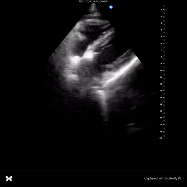
Chest pain is a frequently encountered chief complaint in the Emergency Department and entails a broad differential. Point-of-care ultrasound (POCUS) can be utilized to guide diagnostic decision making and initial triaging. Takotsubo cardiomyopathy presents similarly to acute coronary syndrome and has characteristic findings on echocardiogram. This case presentation details a scenario of ST segment elevation on electrocardiogram and elevated high sensitivity troponin levels, worrisome for a ST elevation myocardial infarction (STEMI). Apical hypokinesis to akinesis and apical ballooning were appreciated on echocardiogram, raising suspicion for Takotsubo cardiomyopathy, subsequently confirmed by coronary angiogram. A cardiac focused point-of-care ultrasound assessment can provide valuable information to aid in diagnostic accuracy.
Case Presentation
A 72-year-old woman with a known history of chronic obstructive pulmonary disease (COPD) presented to the hospital for progressively worsening dyspnea in the previous few days along with new onset chest discomfort in the past one day. Patient was found to have an oxygen saturation of 87% on room air, pH of 7.25 and a pCO2 of 98 on venous blood gas, and was admitted for acute on chronic hypoxic and hypercapnic respiratory failure in the setting of a COPD exacerbation. Patient was intubated for respiratory distress and worsening acuteencephalopathy. Chest radiograph was grossly unremarkable for consolidations or
opacities. A bedside point-of-care ultrasound (POCUS) assessment revealed clear lung zones bilaterally without apparent B lines; however, minimal pleural sliding was appreciated on the left anterior lung zones. Cardiac focused assessment identified marked hypokinesis to akinesis of the entire mid-distal left ventricle with apical ballooning, raising the suspicion of Takotsubo cardiomyopathy (Videos 1-2).

Video 1. Subcostal view with identification of a hyperkinetic basal segment and hypokinetic apex. Apical ballooning is also clearly identifiable in this view. (Click here to view the video in a separate window)

Video 2. Parasternal short axis identifying a hyperkinetic basal segment near the level of the mitral valve with subsequent hypokinetic apical view. The image plane is being panned from base to apex and back. (Click here to view the video in a separate window).
High sensitivity troponin level was elevated at 42 ng/L with an increase to 540 ng/L on repeat testing. Electrocardiogram (ECG) was initially grossly unremarkable for signs of acute ischemic changes, however, repeat ECG revealed ST elevation in the anterior leads. The patient was taken urgently to the catheterization lab where intervention identified mild non-obstructive disease in a right dominant circulation and the diagnosis of Takotsubo cardiomyopathy was confirmed.
Discussion
Chest pain is among the most common chief complaints of patients presenting to the Emergency Department. The differential diagnoses of chest pain remain broad which includes a variety of pathological processes. POCUS has emerged as an indispensable tool for diagnostic accuracy and for aid with initial triaging before considering further confirmatory testing. An emerging consideration is its utility in the acute setting, specifically when trying to differentiate between cardiac and non-cardiac chest pain. Comprehensive echocardiography, usually completed in a formal setting upon request, provides valuable information that can be indicative of ischemic states, including regional wall motion abnormalities, decreased systolic movement, decreased myocardial thickening, valvular function abnormalities, inter-ventricular shunts, and acute papillary muscle dysfunction (1). Alternatively, bedside POCUS in acute settings for assessment of cardiac function and structural abnormalities provides timely objective data but holds greater limitations mainly due to inferior ultrasound quality, variable operator skillsets, and time constraints. of
In our case, we utilized POCUS in an unresponsive, intubated patient, noting discrete regions of hypokinesis-akinesis the left ventricle with apical ballooning, prior to ECG showing elevated ST segments in the anterior leads and a rising troponin level on serial lab tests. Our initial impression based on the POCUS findings was concerning for Takotsubo cardiomyopathy. Given the urgency of the troponin and ECG abnormalities, a Code STEMI was called. Cardiology urgently took the patient to the catheterization lab which confirmed the diagnosis of Takotsubo cardiomyopathy after identifying no obstructive coronary artery disease.
Takotsubo cardiomyopathy often presents very similarly to acute coronary syndrome with elevated markers of myocardial ischemia and ST changes on ECG (2). Hallmarks of this clinical entity include apical hypokinesia and basal segment hyperkinesia on echocardiogram and no obstructive coronary artery disease on coronary angiography. Given the acuity of these findings, this case presentation portrays the importance of utilizing a cardiac focused POCUS assessment to help tailor differential diagnoses and raise index of suspicion not only to acute coronary syndromes, but also to mimicking clinical diseases.
References
- Leischik R, Dworrak B, Sanchis-Gomar F, Lucia A, Buck T, Erbel R. Echocardiographic assessment of myocardial ischemia. Ann Transl Med. 2016 Jul;4(13):259. [CrossRef] [PubMed]
- Prasad A, Lerman A, Rihal CS. Apical ballooning syndrome (Tako-Tsubo or stress cardiomyopathy): a mimic of acute myocardial infarction. Am Heart J. 2008 Mar;155(3):408-17. [CrossRef] [PubMed]
Ultrasound for Critical Care Physicians: Ghost in the Machine
Ross Davidson, DO
Michel Boivin, MD
Division of Pulmonary, Critical Care and Sleep Medicine
University of New Mexico School of Medicine
Albuquerque, NM USA
A 53-year-old woman presented to the emergency department after a sudden cardiac arrest at home. The patient had a history of asthma and tracheal stenosis and had progressive shortness of breath over the previous days. The patient’s family noticed a “thump” sound from the patient’s room, and found her apneic. They called 911 and began cardiopulmonary resuscitation. Paramedics arrived on the scene, found an initial rhythm of pulseless electrical activity. The patient eventually achieved return of spontaneous circulation and was transported to the hospital. On arrival the patient was in normal sinus rhythm, with a heart rate of 110 beats per minute. Blood pressure was 80/45 mmHg, on an epinephrine infusion. The patient was afebrile, endotracheally intubated, unresponsive and ventilated at 30 breaths per minute. An initial chest radiograph was compatible with aspiration pneumonitis and a small pneumothorax. Initial electrocardiogram on arrival had 1mm ST-segment depressions in leads V4 to V6. Transthoracic echocardiography was unsuccessful due to patient’s habitus and mechanical ventilation. Because of the patient’s hemodynamic instability and unknown cause of cardiac arrest, an urgent trans-esophageal echocardiogram (TEE) was performed (Videos 1-3).

Video 1. Mid-esophageal 4-chamber view of the heart.

Video 2. Upper esophageal long-axis view of the pulmonary artery and short axis view of the ascending aorta.

Video 3. Upper esophageal short axis view of the pulmonary artery with the ascending aorta in long axis.
Based on the images presented what do you suspect is the etiology of the patient’s cardiac arrest? (Click on the correct answer for an explanation-no penalty for guessing, you can go back and try again)
Cite as: Davidson R, Boivin M. Ultrasound for critical care physicians: ghost in the machine. Southwest J Pulm Crit Care. 2018;16(2):76-80. doi: https://doi.org/10.13175/swjpcc027-18 PDF
Ultrasound for Critical Care Physicians: Now My Heart Is Still Somewhat Full
Krystal Chan, MD
Bilal Jalil, MD
Department of Internal Medicine
University of New Mexico School of Medicine
Albuquerque, NM USA
A 48-year-old man with a history of hypertension, intravenous drug abuse, hepatitis C, and cirrhosis presented with 1 day of melena and hematemesis. While in the Emergency Department, the patient was witnessed to have approximately 700 mL of hematemesis with tachycardia and hypotension. The patient was admitted to the Medical Intensive Care Unit for hypotension secondary to acute blood loss. He was found to have a decreased hemoglobin, elevated international normalized ratio (INR), and sinus tachycardia. A bedside echocardiogram was performed.

Figure 1. Apical four chamber view of the heart.

Figure 2. Longitudinal view of the inferior vena cava entering into the right atrium.
What is the best explanation for the echocardiographic findings shown above? (Click on the correct answer for an explanation and discussion)
Cite as: Chan K, Jalil B. Ultrasound for critical care physicians: now my heart is still somewhat full. Southwest J Pulm Crit Care. 2016;12(6):236-9. doi: http://dx.doi.org/10.13175/swjpcc054-16 PDF
June 2016 Critical Care Case of the Month
Theodore Loftsgard APRN, ACNP
Julia Terk PA-C
Lauren Trapp PA-C
Bhargavi Gali MD
Department of Anesthesiology
Mayo Clinic Minnesota
Rochester, MN USA
Critical Care Case of the Month CME Information
Members of the Arizona, New Mexico, Colorado and California Thoracic Societies and the Mayo Clinic are able to receive 0.25 AMA PRA Category 1 Credits™ for each case they complete. Completion of an evaluation form is required to receive credit and a link is provided on the last panel of the activity.
0.25 AMA PRA Category 1 Credit(s)™
Estimated time to complete this activity: 0.25 hours
Lead Author(s): Theodore Loftsgard, APRN, ACNP. All Faculty, CME Planning Committee Members, and the CME Office Reviewers have disclosed that they do not have any relevant financial relationships with commercial interests that would constitute a conflict of interest concerning this CME activity.
Learning Objectives:
As a result of this activity I will be better able to:
- Correctly interpret and identify clinical practices supported by the highest quality available evidence.
- Will be better able to establsh the optimal evaluation leading to a correct diagnosis for patients with pulmonary, critical care and sleep disorders.
- Will improve the translation of the most current clinical information into the delivery of high quality care for patients.
- Will integrate new treatment options in discussing available treatment alternatives for patients with pulmonary, critical care and sleep related disorders.
Learning Format: Case-based, interactive online course, including mandatory assessment questions (number of questions varies by case). Please also read the Technical Requirements.
CME Sponsor: University of Arizona College of Medicine
Current Approval Period: January 1, 2015-December 31, 2016
Financial Support Received: None
History of Present Illness
A 64-year-old man underwent three vessel coronary artery bypass grafting (CABG). His intraoperative and postoperative course was remarkable other than transient atrial fibrillation postoperatively for which he was anticoagulated and incisional chest pain which was treated with ibuprofen. He was discharged on post-operative day 5. However, he presented to an outside emergency department two days later with chest pain which had been present since discharge but had intensified.
PMH, SH, and FH
He had the following past medical problems noted:
- Coronary artery disease
- Coronary artery aneurysm and thrombus of the left circumflex artery
- Dyslipidemia
- Hypertension
- Obstructive sleep apnea, on CPAP
- Prostate cancer, status post radical prostatectomy penile prosthesis
He had been a heavy cigarette smoker but had recently quit. Family history was noncontributory.
Physical Examination
His physical examination was unremarkable at that time other than changes consistent with his recent CABG.
Which of the following are appropriate at this time? (Click on the correct answer to proceed to the second of four panels)
Cite as: Loftsgard T, Terk J, Trapp L, Gali B. June 2016 critical care case of the month. Southwest J Pulm Criti Care. 2016 Jun:12(6):212-5. doi: http://dx.doi.org/10.13175/swjpcc043-16 PDF